Mitosis Duration
Mitosis Duration
Although the cell cycle duration may vary significantly, the duration of mitosis is often short and rather consistent. Different stimuli, including chemicals and other environmental factors, may affect the mitosis duration. Based on cell morphology, the duration of mitosis for individual cells can be determined.

The HoloMonitor cell tracking tool allows determination of the mitosis duration time. Each colored line corresponds to one mitotic cell. The graph on the top shows controls, while the graph below shows cells treated with a cytostatic agent. The treatment clearly prolonged the mitosis duration.
Mitosis Duration using HoloMonitor®
The HoloMonitor cell tracking tool allows determination of mitosis duration for individual cells as selected by the user. Duration of mitosis is determined without the use of labels or stains, and based on live-cell analysis providing both images and quantitative data. Examples of output:
- Time-lapse images and videos visualizing the various morphological changes cells undergo during mitosis.
- Total mitosis time for each individual cell.
- Graphs and data showing when each cell was in mitosis.
Assessment of mitosis duration can be complemented with other analyses like cell proliferation, cell motility and migration and/or evaluation of morphological parameters.
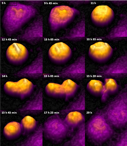
Cell division studied with holography. From the first rounding up of the cell to completed cytokinesis is approximately 4 hours.
- PHI HoloMonitor M4 with Hstudio software
- Culture vessels and PHI vessel holder for selected vessel
- PHI HoloLid for selected vessel