


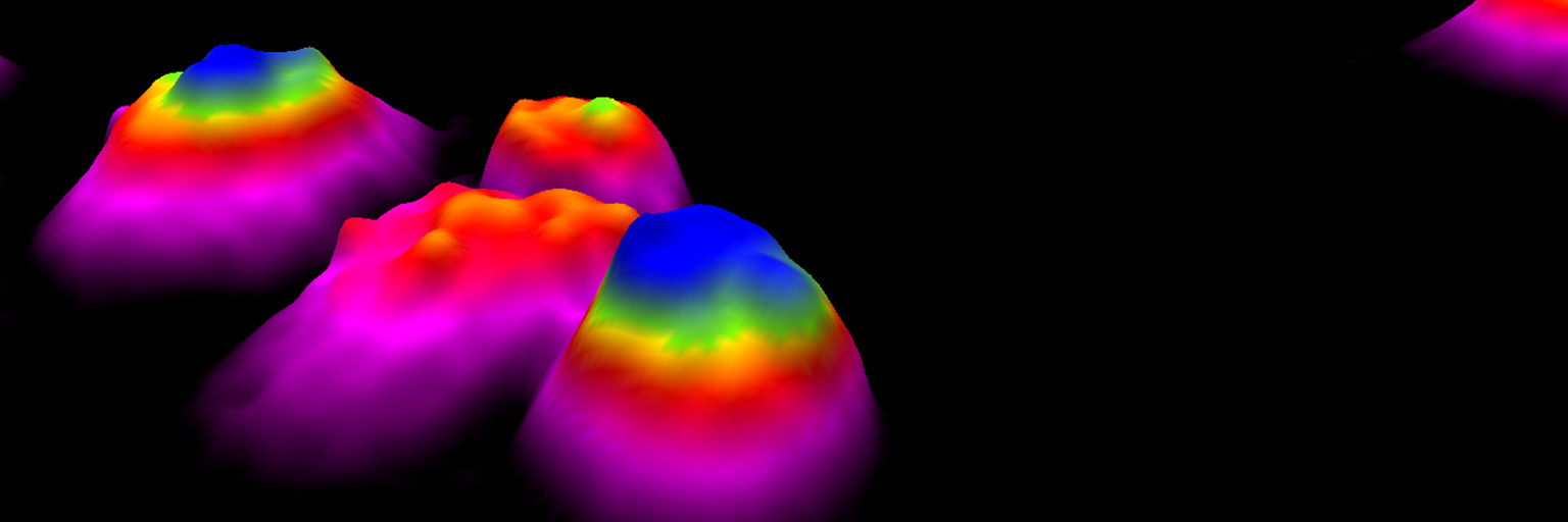
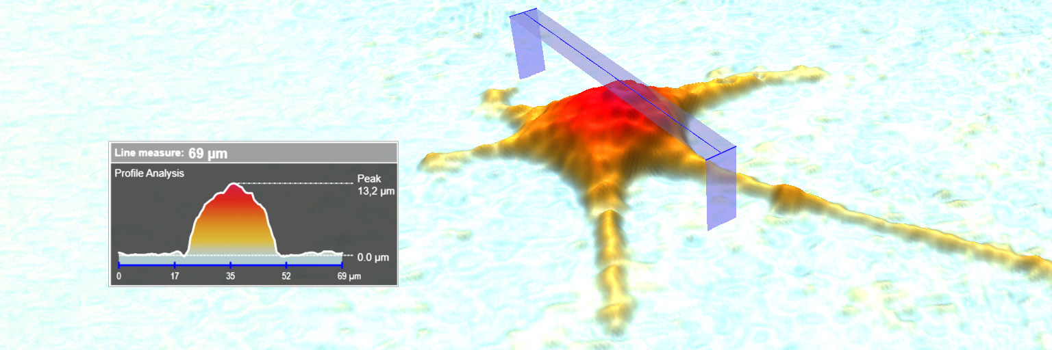
Cell Morphology Assay
The HoloMonitor Cell Morphology Assay enables non-invasive live cell quantification of a wide range of morphological properties, including individual cell volume, area and thickness.
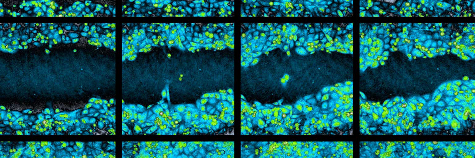
Wound Healing Assay
The HoloMonitor Wound Healing Assay analyze in vitro cell migration. Cell migration speed is automatically determined, directly in your incubator.
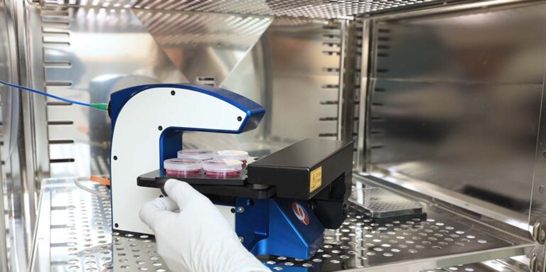
Single-cell Analysis Assays
WIth single-cell analysis assays from PHI, you can study cell movement and morphology of living cells directly inside your incubator.