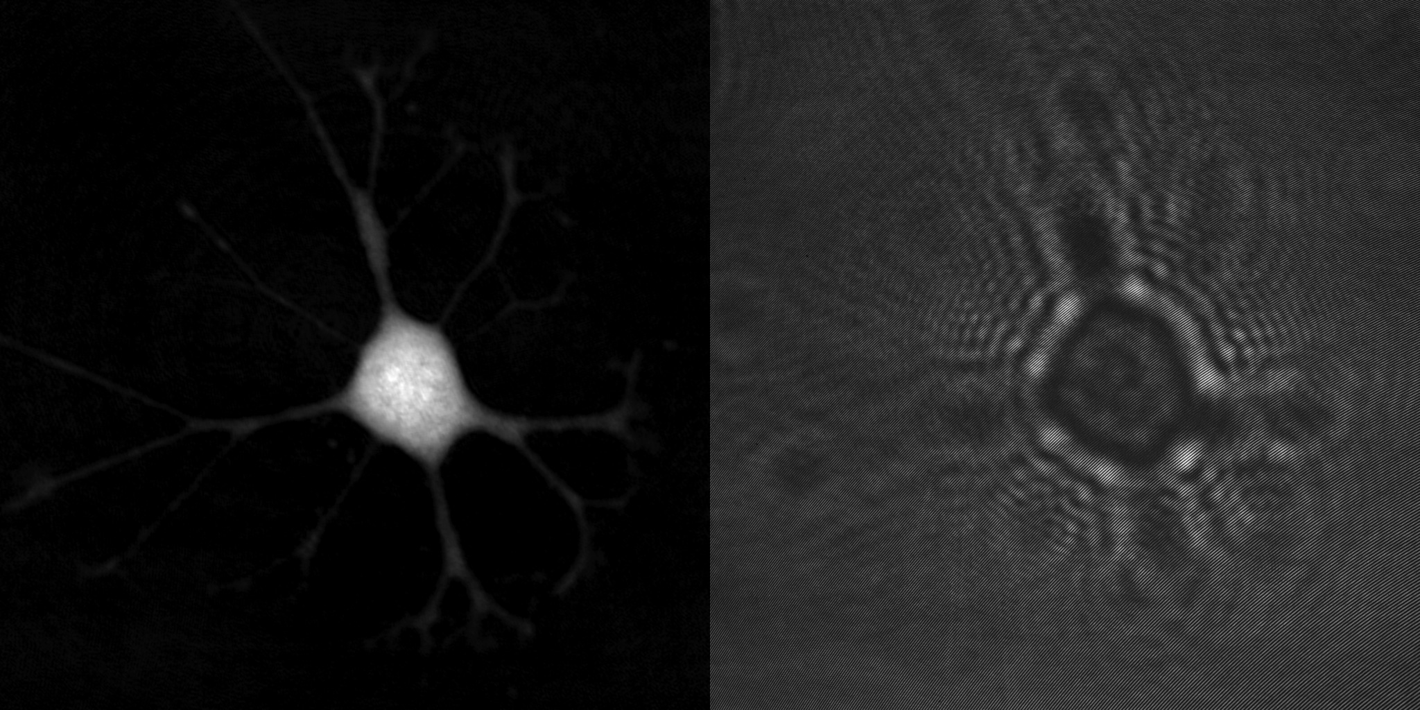
A Time-lapse, Label-free, Quantitative Phase Imaging Study of Dormant and Active Human Cancer Cells
Journal: JoVe (video journal) (2018)
Research Areas: Cancer Research
Cell Lines: KHOS
Learn more …

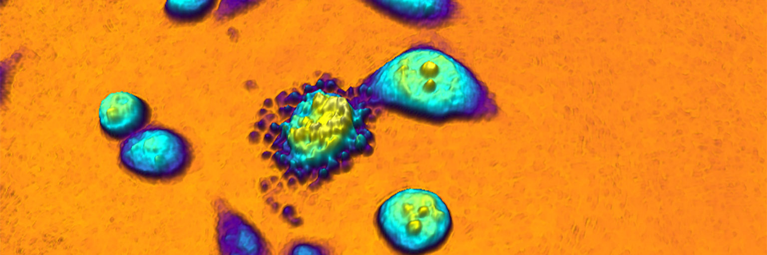
Applications of Label-free, Quantitative Phase Holographic Imaging Cytometry to the Development of Multi-specific Nanoscale Pharmaceutical Formulations
Journal: Cytometry Part A 2017 (2017)
Research Areas: Pharmacology
Cell Lines: HeLa, L929
Learn more …