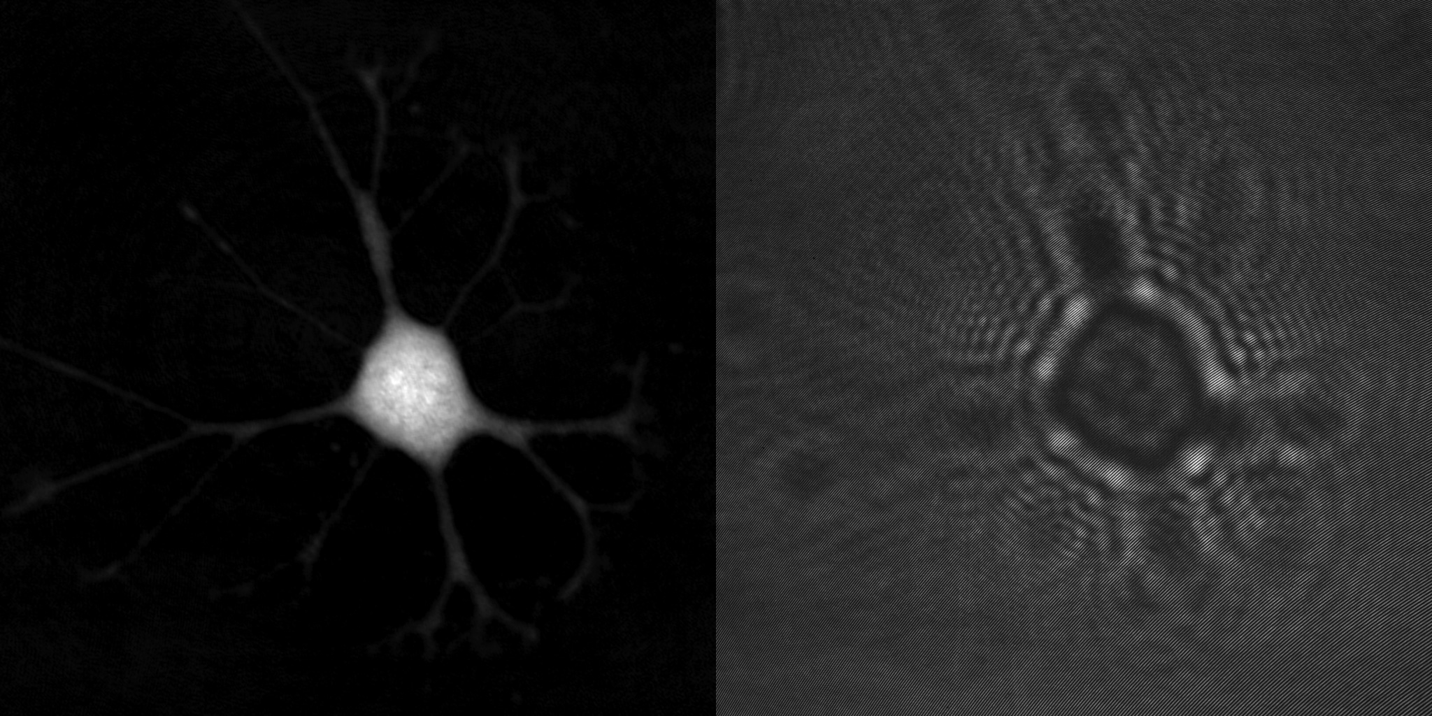
Moving into a New Dimension: Tracking Migrating Cells with Digital Holographic Cytometry in 3D
Journal: Cytometry Part A (2018)
Research Areas: Cancer research
Learn more …
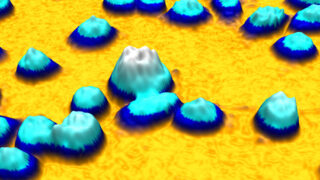
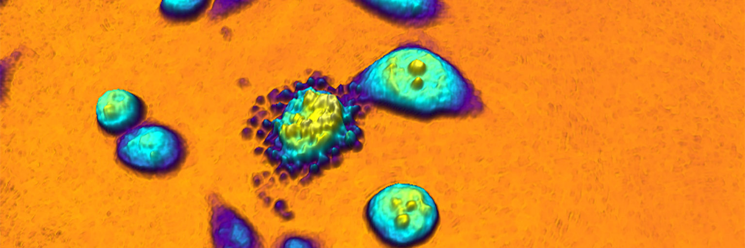
HoloMonitor M4: holographic imaging cytometer for real-time kinetic label-free live-cell analysis of adherent cells
Journal: Proceedings, Quantitative Phase Imaging II (2016)
Research Areas: Method development
Learn more …