Single Cell Tracking Assay
Your non-invasive and automatic single cell tracking assay
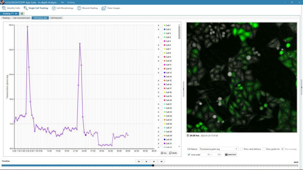
The HoloMonitor® Single Cell Tracking assay offers user-friendly software for continuous, label-free imaging and tracking of adherent cells, enabling in-depth characterization of heterogeneous cell behavior at the single-cell level. With the add-on fluorescence unit, you can add a green fluorescence dimension to the holography data, providing enhanced insights into your cell cultures.
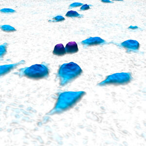
Watch L929 mouse cells in action with the HoloMonitor Single Cell Tracking Assay using label-free holography.
Benefits
Why choose the HoloMonitor Single Cell Tracking Assay?
Non-invasive
There is no need for cellular stains or labels to track the cells, making it perfect for biologically relevant long-term studies.
High-content
Explore individual and population cell behavior over time with further insights from the fluorescence add-on.
Easy-to-use
Get support in your daily lab flows and cell culture work with easy-to-use software.
Assay Output
What results do you get with the label-free Single Cell Tracking Assay?
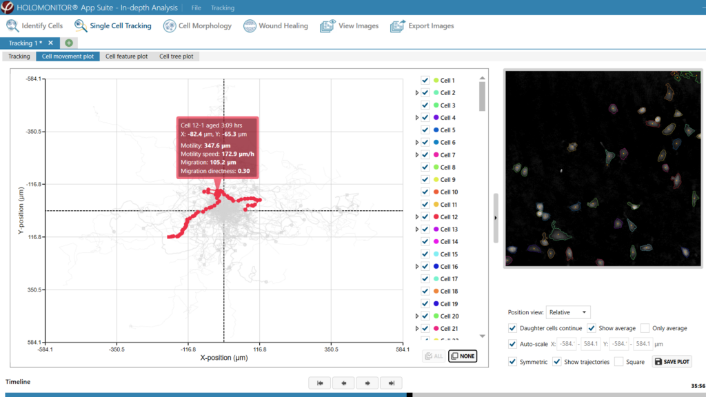
- Cellular movement plot with motility, motility speed, migration and migration directness data
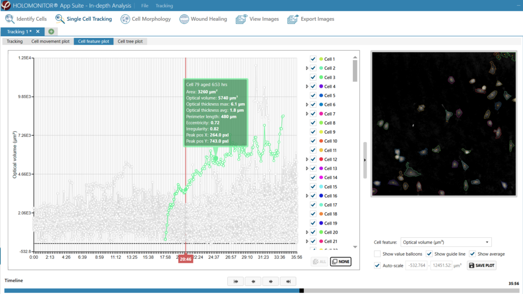
- Cell feature plot with detailed kinetic morphology data
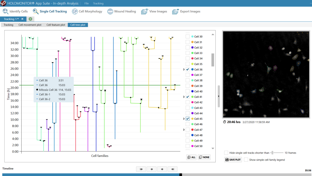
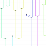
- Cell tree plot of all tracked cells and their cell family
- Time-lapse images and videos
Time-Efficient.
Powerful algorithms immediately generate automatic tracking results
Our development team thrives by increasing user productivity. Therefore, the Single Cell Tracking Assay is optimized to boost your analysis efficiency. It automatically tracks all cells in the field of view when imaging your cell sample and presents your images and results in a clear and graphic way.
Like all HoloMonitor live cell assays, you set up your label-free assay in the same easy and straightforward way in the software and get guided through the experiment workflow. Firstly, this not only immensely reduces your hands-on lab time but also increases the reproducibility of your assay. Consequently, this will allow you to achieve more reliable and relevant results in your cell-based research.
- Guided assay workflows reduce your hands-on lab time and increase reproducibility
- Powerful algorithms immediately generate automatic tracking results
- Export your data for further investigation
On-demand Webinar
How label-free single-cell tracking reveals unique cell behavior
Learn how the HoloMonitor Single Cell Tracking Assay facilitates long-term imaging and tracking of adherent cells, allowing label-free analysis of heterogeneous cell behavior on a single-cell level.
Key Applications
What can you study with HoloMonitor Single Cell Tracking Assay?
Collective migration studies
Cellular heterogeneity studies
Drug discovery & development
Cell cycle studies
Single-cell analysis
Cell families
Kinetic cell morphology studies
Co-culture
Cell death
Transfection efficiency
Uptake assay
Reporter gene expression
Non-invasive.
A cell-friendly tool for tracking live cell cultures in real time with the option for additional fluorescence insights
Like all HoloMonitor live cell assays, it lets you follow cells over time in a non-invasive way. In fact, no cytotoxic stains, markers, or cellular labels are needed to track the cells. This lets you keep light exposure to a minimum and reduce photocytotoxicity when deciding to add fluorescence for additional insights. Label-free assays not only reduce the risk of unwanted toxicity. They also allow you to reuse your cell samples, as you do not have to add reagents to the sample. Hence making it possible for you to save rare and precious cells, such as stem cells and primary cells.
Most importantly, you are able to follow everyday cell life right inside the incubator. By imaging inside an incubator, HoloMonitor creates a stable and relevant assay environment for your cells. Therefore, the HoloMonitor Single Cell Tracking assay is ideal for long-term studies of your live cell cultures.
- No cytotoxic stains are needed to track your cells
- Imaging right inside the incubator
- Perfect for biologically relevant long-term studies
Single Cell Tracking Assay Protocol
How do you set up this Single Cell Tracking assay?
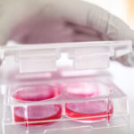
1. Sample preparation
- Seed label-free adherent cells in your standard cell culture vessel.
- Use HoloLids for best image quality and data acquisition.
- Then, place the cell culture vessel in the incubator onto the HoloMonitor motorized stage.
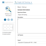
2. Assay setup
- Select the HoloMonitor Single Cell Tracking Assay in the software. Next, name your experiment and define treatments.
- Then, selecting wells, imaging positions, duration, and interval. Start imaging and walk away!
3. Explore results
- Get detailed cell movement and kinetic morphology data, as well as entire cell family trees. Analyze the experiment with other live cell assays for further studies.
- Export the raw data for further analysis. And re-use your cells for other experiments!
High-content.
Get more data on every single cell within a population
All our HoloMonitor live cell assays are developed together with cell biologists since we want to make sure that they won’t waste your limited lab time and resources. On the contrary to the traditional way of working, where you had to set up several different time- and cell-consuming assays for each assay result you wish to get. You can re-use imaging results from one experiment and easily analyze them in multiple other HoloMonitor assays — from home if you have to.
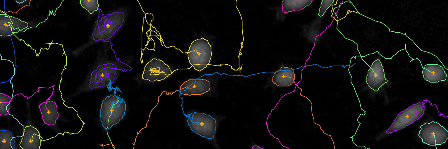
The Single Cell Tracking Assay lets you tap into the immense potential resulting from multiple automatic assay results. Specifically, making it possible for you to explore and combine cell morphology and cell movement data. As a result, you get to study over 30 label-free cell morphology characteristics with the option to add a green fluorescence dimension with the add-on fluorescence unit. The outstanding individual cell tracking of the system is loved by our users. In short, you get to follow in detailed movement plots single cells and cell populations in their migration and motility behavior.
- More insights in one experiment setup
- Study 30+ morphology features from one experiment
- Follow in advanced movement plots both single cells and their entire cell families
Study heterogeneous cell behavior
The quantitative holographic 3D images created by HoloMonitor allow you to study cell populations on a single-cell level without the need for labels or stains. As a result, you are able to get insights into single cells and their entire family tree in a non-invasive way while not compromising on data. However, the HoloMonitor add-on fluorescence unit allows adding a green fluorescence dimension to the label-free holography data and studying your cells’ behavior in even more detail.
Mammalian cells can be highly dynamic in both morphology and behavior. Characterizing and tracking diverse cell behavior over time on a single-cell level is critically important when studying special or rare events. As an example, you can characterize cells as becoming drug-resistant or going through a transition event, such as cell differentiation. Hence, by tracking every single cell within a population, you never miss a cellular event that you are interested in. Moreover, using HoloMonitor live cell assays, you study your cells in real-time, over long periods of time — basically, you have a 24/7 live-cell TV right inside your incubator.
- Get insights both on individual cells and entire family trees
- Follow everyday cell life, such as cell division, cell differentiation, cell migration, and cell death
- Never miss a cellular event with 24/7 continuous live cell imaging
Combine wound healing with HoloMonitor Single Cell Tracking Assay
Add to your wound healing results with single cell tracking for more kinetic data.
Easily export all your data for further analysis
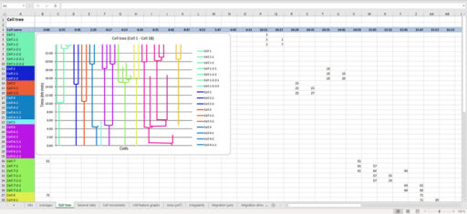
You can export all the data used for the HoloMonitor Single Cell Tracking analysis to an XLSX file. Hence, you can use Excel to open and analyze the data further with your own methods.
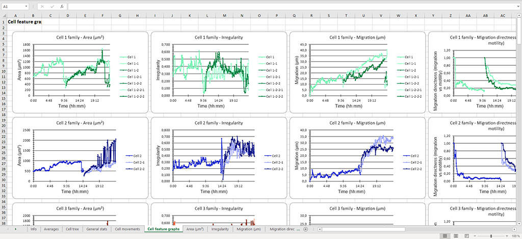
The export contains raw data for more than 30 morphological features, such as cell area, volume, thickness, but also cell migration and motility. Hence, you can extract the data that is relevant to you and make your own analysis and result presentation of, for example, cell cycle length.
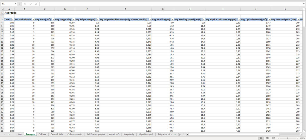
However, you also get pre-made graphs for each analyzed cell family showing all cell features, movement plots, and cell trees. These give you a useful overview of your results and helps you pick out interesting cells to analyze further.
Easy-to-use.
Modern software with a guided workflow
The Single Cell Tracking Assay is designed to meet the current demands of cell researchers. To help you to succeed in your research, we continuously develop our assays to adapt to your future needs.
At any time, you can swiftly re-analyze recorded time-lapse images with another HoloMonitor live cell assay. You can come back to your experiment data at a later point and even work on your data analysis from home. Therefore, you can quickly obtain multiple results from the same cell sample and experiment setup — saving you time, money, and precious cells.
Combined insights of both morphology and movement behavior of cells are absolutely a powerful tool when studying complex biological questions such as EMT in cancer metastasis or stem cell differentiation kinetics. However, it is not just kinetic morphology and movement data you can generate from the same original tracking data set. In fact, you can study many events in everyday cell life using the same tracking results. For example, you can explore cell proliferation, cytotoxicity, or drug responses.
- Cell imaging and analysis software for biologists, by biologists
- Get more results from multiple assays in the software and expand your insights
- Study other cellular events using the same experiment results, such as cell proliferation, cytotoxicity, or drug response
Single Cell Tracking Assay FAQ
The HoloMonitor system uses quantitative phase imaging to track both the movement and morphology of adherent cells. It monitors and analyzes the cells over extended periods of time, with no need for labels or genetic modifications of any kind.
What is the HoloMonitor Single Cell Tracking Assay?
The HoloMonitor Single Cell Tracking Assay is the most advanced and powerful feature of the HoloMonitor live cell imaging system. It lets you track and classify single cells in heterogeneous cell cultures, all label-free. You get both motion and kinetic morphology data for each individual cell and mean values for all cells included in the analysis.
Cell movement can be categorized into non-directional cell motility and directional cell migration. In short, cell motility is the random cell movement, which is present in almost every cell culture. On the other hand, cell migration is cell movement that is not random but rather caused by a cell attractant or repellent.![]()
No! In addition to cell motion, HoloMonitor measures a wide range of morphological properties of the tracked cells. For instance, cell volume, cell area, cell thickness, cell irregularity, and cell roughness are all automatically measured throughout the experiment.
Of course not! The HoloMonitor App Suite software lets you plot-relevant features to monitor how they change over time and as your cells divide. However, you can also export all raw data along with pre-made graphs, movement plots, and cell trees to Microsoft Excel. Here, you can analyze and present the results in the way you want.

The beauty is in the single-cell analysis, the microscope creates amazing time-lapse movies, and it is very easy to set up for time-lapse imaging. Also, the price is amazingly low. Every lab should have one!
Stina Oredsson, PhD
Lund University
Featured Publications
Browse our publication library for more references and see how our worldwide HoloMonitor users run their single-cell tracking assays.

Bioactive Phenolics from Vinegar–Egg Accelerates Acute Wound Healing by Activation of Focal Adhesion and Mitogen-Activated Protein Kinase Signaling
Journal: Nutrients (2025)
Research Areas: Cell Research
Cell Lines: MEF

Dissecting Morphological and Functional Dynamics of Non-Tumorigenic and Triple-Negative Breast Cancer Cell Lines Using PCA and t-SNE Analysis
Journal: Cancer Reports (2025)
Research Areas: Cancer Research
Cell Lines: MCF10A, MDA-MB-231
TNIK Regulates Cytoskeletal Organization to Promote Focal Adhesion Turnover and Mitosis in Lung Adenocarcinoma
Journal: Frontiers in Bioscience-Landmark (2025)
Research Areas: Cancer Research
Cell Lines: MDA‑MB‑231, MCF‑7

Novel Low-Cytotoxic and Highly Efficient Type I Photoinitiators for Visible LED-/Sunlight-Induced Photopolymerization and High-Precision 3D Printing
Journal: Angewandte Chemie International Edition (2025)
Research Areas: Molecular chemistry
Cell Lines: C3H10 T1/2