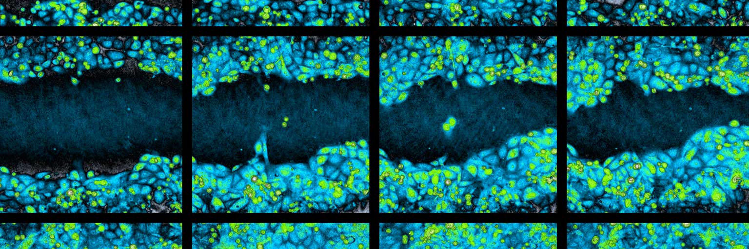
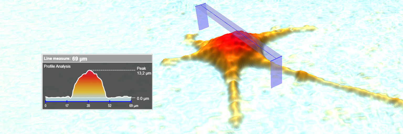
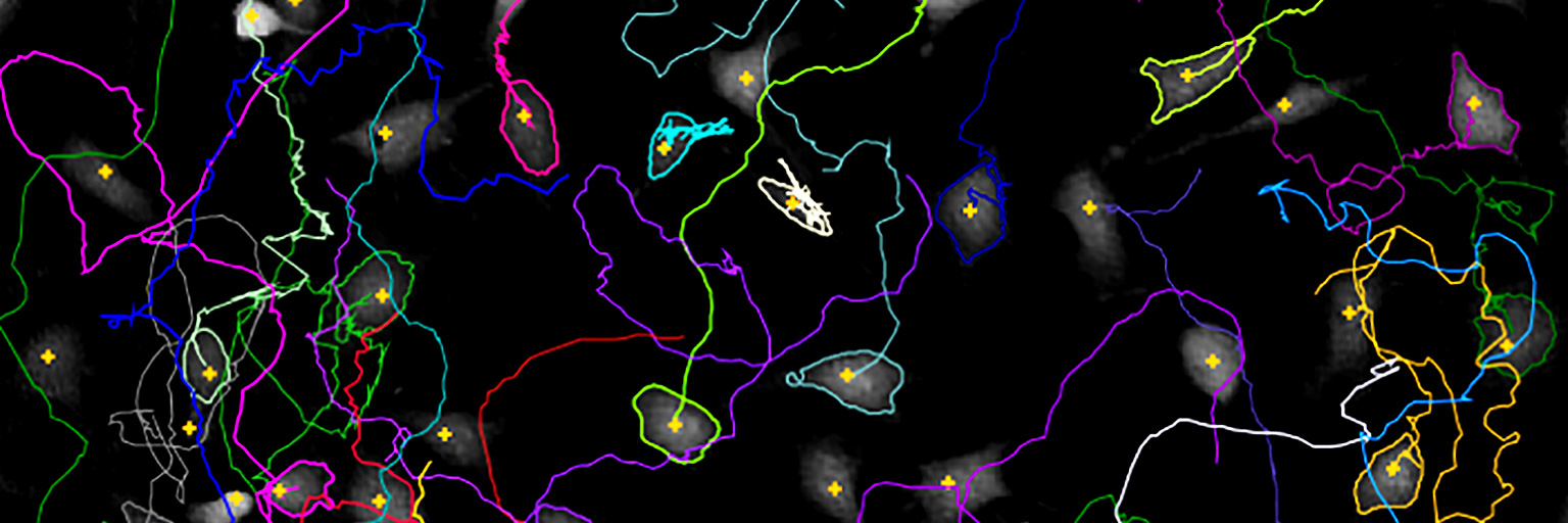
created using HoloMonitor®
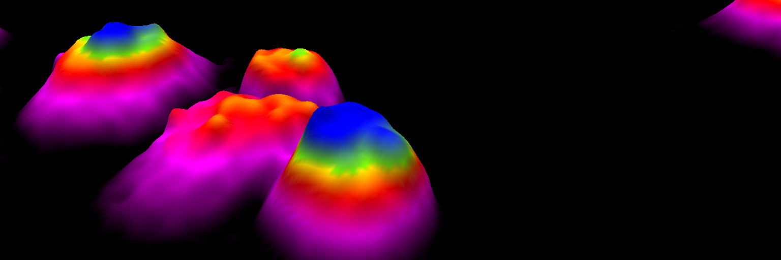

Live Cell Morphology Assay
The label-free HoloMonitor® Cell Morphology Assay enables non-invasive live cell imaging and analysis of a wide range of morphological properties, including individual cell volume, area and thickness.
Individual Cell Morphology
Morphological changes caused by treatment or cellular transformation occur early, making kinetic cell morphology studies a significantly more sensitive tool than indirect biochemical assays to observe cellular response.
In the past, in vitro cell morphology has been difficult and time-consuming to quantify as it involves inconsistent staining and handling of complicated cell imaging software. Contrary to conventional live cell imagers, HoloMonitor acquires a time-lapse sequence of label-free quantitative phase images.
Cell-friendly Imaging
Since no phototoxic or fading fluorescent labels are required, an unlimited number of label-free and cell-friendly time-lapse images can be acquired just minutes apart. This allows HoloMonitor to non-invasively quantify treatment response of adherent cell populations, by automatically quantifying alterations in individual cell morphology.
Cell Morphology Analysis — Scatter Plots & Histograms
The HoloMonitor Cell Morphology Assay measures a wide range of morphological properties, including cell volume, area and thickness. Scatter plots help visualize differences between control and treated cells or to compare cellular characteristics at different time-points of the experiment.
Gating regions enable classification in subpopulations and identification of (rare) cells of interest, for further studies using HoloMonitor Cell Tracking. Histograms show cell population distribution of selected morphological parameter at selected time-points.
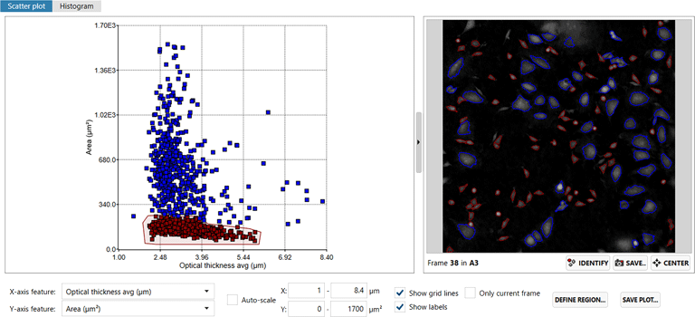
Screenshot of the HoloMonitor Cell Morphology Assay showing cells classified in subpopulations using gating regions.
Label-free Live Cell Assays
HoloMonitor offers several label-free live cell assays, ranging from cell culture quality control to single-cell tracking. After time-lapse image sequences have been recorded at selected sample positions, each of the assays may be used to provide additional results, without requiring further experiments or lab work.
For example, cell motility may be quantified together with cell morphology by tracking cells individually or by applying the HoloMonitor Cell Motility Assay, on the acquired time-lapse image sequence. As the saved image sequence is reused, no further samples are necessary. Additionally, cell proliferation data may also be generated by applying the HoloMonitor Cell Proliferation Assay, again using the previously acquired and saved image sequence.

The HoloMonitor® Live Cell Assays
(screenshot of the HoloMonitor App Suite software)
Jing Huang et al.

Some of the morphological properties that can be analyzed using HoloMonitor.
Links to further applications based on cell morphology analysis
Mitosis Duration
- Total mitosis time for each individual cell.
- Graphs and data showing when each cell was in mitosis.
Cell Cycle Analysis
- Assess cell cycle duration time.
- Detect drug effects on cell cycle phase distribution.
Cell Death Kinetics
- Detect early signs of drug responses before the actual cell death occurs.
- Identify dying cells.
HoloMonitor® Kinetic Morphology Assessment
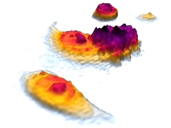
A vast line of morphological parameters can be determined during a time-lapse experiment using HoloMonitor. Like all HoloMonitor applications, morphology assessment is cell-friendly and analysed without the use of any labels or stains. Morphological parameters are, for example, useful
- For evaluation of cell culture health and quality
- As early and sensitive markers to detect drug effects
- To provide information on drug mode of action
- For identification of dying cells
Cell morphology analysis is often complemented by cell proliferation and cell motility studies to provide a complete picture.
References

Quantitative evaluation of morphological changes in activated platelets in vitro using digital holographic microscopy
Authors: Yutaka Kitamura et al.
Journal: Micron (2018)
Research Areas: Method development
Cell Lines: Human platelets
Keywords: HoloMonitor M4, Cell morphology, Digital holographic microscopy, Platelets, Aggregation, Activation

A Time-lapse, Label-free, Quantitative Phase Imaging Study of Dormant and Active Human Cancer Cells
Authors: Jing Huang et al.
Journal: JoVe (video journal) (2018)
Research Areas: Cancer Research
Cell Lines: KHOS
Keywords: HoloMonitor M4, Cell morphology, Cell motility, Cell migration, Cell proliferation
