Wound Healing Assay | Scratch Assay
Your automated and non-invasive wound healing assay
The label-free HoloMonitor® Wound Healing Scratch Assay enables cell-friendly, non-invasive cell migration analysis. It continuously images your samples, identifies every cell, and automatically analyzes the results. As a result, you receive accurate, kinetic gap closure data in real-time. In addition, combined with Culture-Inserts from ibidi®, you achieve superior reproducibility and consistency between your wound healing experiments.
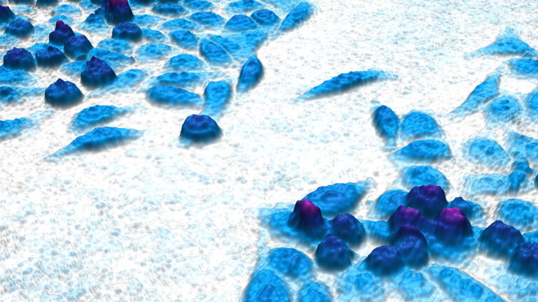
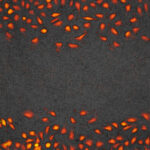
Time-lapse video of a closing wound healing gap, captured with HoloMonitor.
Benefits
Why choose the HoloMonitor Wound Healing Scratch Assay?
Accurate
Get exact cell segmentation with digital holography to measure what you actually want to study.
Non-invasive
Avoid known interferences from cellular markers and achieve biologically relevant results.
User-friendly
Save time with the automated and guided App Suite software, designed by and for cell biologists.
Reproducible
Combine this assay with the ibidi Culture-Inserts for an easy and consistent experimental setup.
Assay Output
What results do you get with the HoloMonitor Wound Healing Scratch Assay?
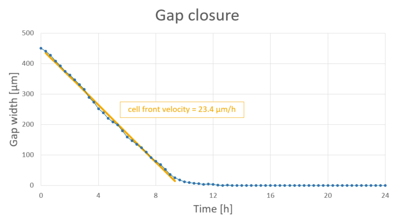
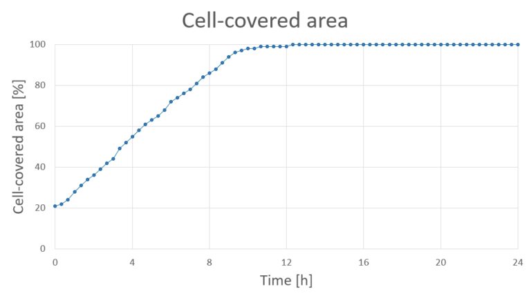
The HoloMonitor Wound Healing Assay automatically determines your gap closure kinetics in real-time. You can follow the process in a time-lapse video and the software will provide you with the relevant data for each time point.
- Kinetic gap closure data over time
- Gap width [µm]
- Cell-covered / cell-free area [µm², %]
- Cell front velocity [µm/h]
- Images and time-lapse videos of the gap closure process
- Combine with the Single Cell Tracking Assay and Cell Morphology Assay for deeper insights
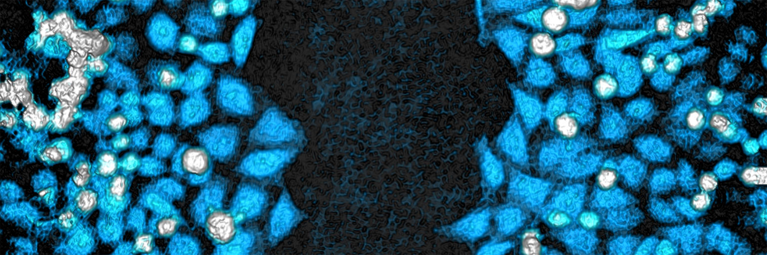
Label-free time-lapse video showing non-treated human breast cancer cells (JIMT-1) closing a wound healing gap.
Accurate measurements.
Exact cell segmentation measures what you actually want to study
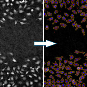
Compared to traditional phase contrast microscopy, the quantitative phase imaging technology used by HoloMonitor offers superior cell segmentation and identification. As a result, you get a distinct separation between cells and the background. This enables the HoloMonitor software to provide accurate data, in 3D, on every single cell. Therefore, using HoloMonitor, you can be sure to measure what you actually want to study.
On-demand Webinar
Watch our Wound healing assay webinar together with ibidi!
This webinar showcases how you how to run wound healing assays that get you biologically relevant and reproducible data using the HoloMonitor Wound Healing Assay protocol.
Key Applications
What can you study with the HoloMonitor Wound Healing Assay?
Wound healing (scratch) studies
Transwell studies
Co-culture studies
Cell invasion studies
Cell-friendly wound healing analysis.
Study your cells non-invasively right inside your standard incubator
The non-invasive quantitative phase imaging technology used by HoloMonitor does not affect your cells in any way. In fact, you image your wound healing experiments without any labels or stains. Moreover, the low-energy laser keeps photocytotoxicity at a minimum. In addition, everything takes place in your standard incubator. As a result, by eliminating elements known to influence cellular behavior, you are sure to get biologically relevant results.
Wound Healing Assay Protocol
How do you set up this in vitro wound healing assay?
1. Sample preparation
- Prepare your wound healing cell cultures as you normally would. With ibidi’s Culture-Inserts, you will get a defined gap suitable for HoloMonitor.
- Use HoloLids for optimal image quality and data acquisition.
- Click your cell culture vessel onto the HoloMonitor motorized stage and you are ready to go!
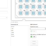
2. Assay setup
- Select the Wound Healing Assay in the software and follow the guided workflow. Next, name your experiment and define treatments.
- Add the positions you want to image. Also, choose how often and for how long you want to image your cells.
- Start imaging and walk away!
3. Explore results
- Receive kinetic gap closure data and time-lapse videos in real time. Analyze the results with other live cell assays for deeper insights into your cells.
- Export the raw data to Excel for further analysis and customized data presentation.
- Re-use the untouched cells for other experiments!
Reproducible setup.
Consistent setup with non-biased data collection and analysis
The HoloMonitor Wound Healing Assay gets you relevant results from controlled wound healing conditions. Firstly, the ibidi Culture-Inserts allow for easy and consistent gap creation. Secondly, HoloMonitor software offers a streamlined software setup. In addition, the whole assay takes place in the stable environment of your standard incubator. Moreover, you can use HoloLids™ to achieve optimal image quality.
Easy-to-use, yet powerful, software.
Save time in your lab with automation
The HoloMonitor App Suite cell imaging software is designed by and for cell biologists. In fact, the user-friendly workflow guides you through the whole wound healing assay, from setup to data presentation. Also, with pre-defined templates for the ibidi Culture-Inserts, you can set up in no time. In addition, the system automatically performs image acquisition, analysis, and data presentation. So you can follow your experiment in real time.
Get more data from your samples
A major benefit with HoloMonitor is that you can get more data from your samples compared to traditional methods. Not only does it leave your cells unaffected. But it also allows you to reanalyze your already collected image sequences in new ways.
Re-use your cells after the imaging is done
Above all, HoloMonitor technology images your cells without affecting them in any way. As a result, they are completely untouched when the experiment is over. This means that you can use the very same cells for subsequent experiments. Therefore, you save both time, money, and cells.
Re-analyze your data in new ways
Moreover, data sets recorded with the Wound Healing Assay can be used for other App Suite analyses as well. For example, if you do a gap closure experiment and realize that you need proliferation or morphology data, you can simply re-analyze the old imaging experiment with another HoloMonitor Assay. In other words, there is no need to redo the experiment. And you will get data on the exact same cells as your original study.
Deeper insights from your wound healing experiment
Certainly, the HoloMonitor Wound Healing Assay can be combined with other HoloMonitor Assays. Therefore, you can get an even better understanding of your experiments.
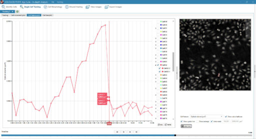
With the Single Cell Tracking Assay, you can track every single cell in your image sequence and get detailed data on cell movement and cell division. This lets you detect unique cell behavior on a single-cell level and reveals not only when, but also how, the gap was closed.
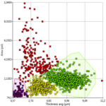
Moreover, you can study over 30 different cellular features with the Cell Morphology Assay. For example, you can explore cell volume or thickness, down to the single-cell level. In addition, the results can be visualized in a dot plot. And this may reveal sub-populations of cells that behave differently in your gap closure experiment.

The advantage of this compared to traditional gap closure analyses is that it is a high throughput system, uses standard culture conditions with an automated image capture, segmentation and tracking of cells and combines functional aspects of cellular behavior with morphological properties on cell level.
Sofia Mogren et al.
Mast cell tryptase enhances wound healing by promoting migration in human bronchial epithelial cells, Cell Adhesion & Migration (2021)
Featured Publications
Browse our publication library for more references and see how our worldwide HoloMonitor users run their wound healing analysis.
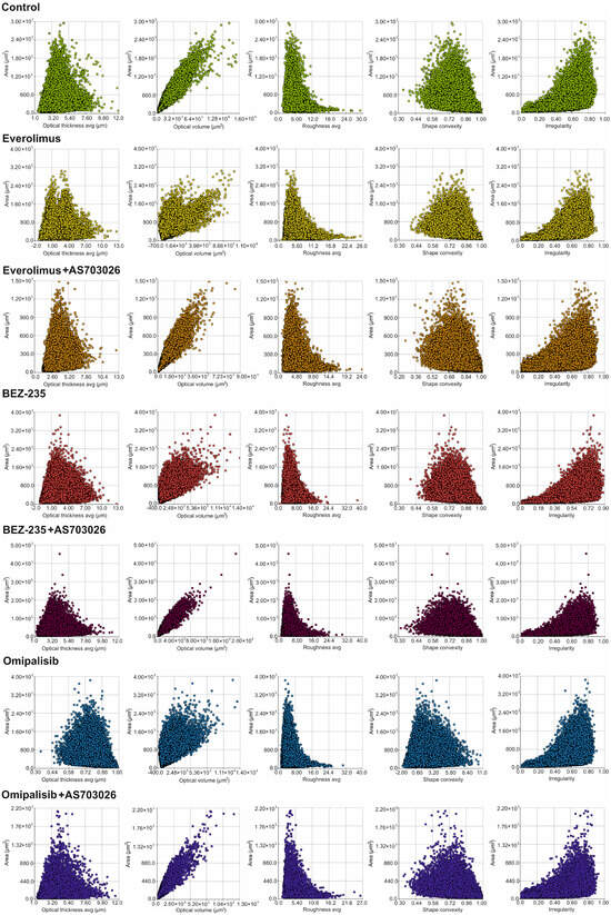
Changes in Melanoma Cell Morphology Following Inhibition of Cell Invasion by Third-Generation mTOR Kinase Inhibitors
Journal: International Journal of Molecular Sciences (2025)
Research Areas: Cancer Research
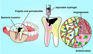
Dual-functional injectable hydrogels as antimicrobial and angiogenic therapeutics for dental pulp regeneration†
Journal: Journal of Materials Chemistry B (2025)
Research Areas: Material chemistry
Cell Lines: SVEC4-10

FOXO3A Plays a Role in Wound Healing by Regulating Fibroblast Mitochondrial Dynamics
Journal: Journal of Investigative Dermatology (2024)
Research Areas: Woundhealing
Cell Lines: Primary fibroblasts

Methods to Investigate Cell Migration
Journal: Cell Migration in Development, Health and Disease (2024)
Research Areas: Cell Biology