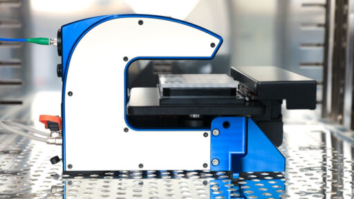
The HoloMonitor® live cell imaging system enables long-term, non-invasive analysis of cell cultures within a standard CO₂ incubator. Therefore, cell biologists worldwide use HoloMonitor to automatically get live single-cell morphology, migration and proliferation data in real-time.
Label-free imaging of living cells means long-term imaging of living cells
The HoloMonitor live cell imaging system uses non-invasive digital holography to monitor your cells. It works completely label-free, uses a low-energy laser, and operates inside your standard incubator. Hence, you can image your cells with a high temporal resolution, for as long as you need, without affecting them. Of course, this is ideal not the least when you work with hard-to-get primary cells. As you do not alter your cells, you can reuse them for other experiments once the imaging is done.
Live cell culture monitoring with HoloMonitor
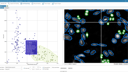
The HoloMonitor live cell imaging system continuously records multiple time-lapse sequences of your cells right inside your incubator. The image sequences contain a multitude of data, with over 30 cellular features recorded for every single cell. Therefore, it non-invasively gives answers to scientific questions that would normally require you to sacrifice your cells. In essence, HoloMonitor lets you do proliferation, motility, dose-response and wound healing studies without wasting any cells.
Multiple experiments with only one sample
Every image taken with HoloMonitor contains all obtainable data, no matter what assay you were doing while recording it. Hence, you can reanalyze any experiment as a new type of assay. So, if you did a motility assay and need proliferation data on the same cells, just reanalyze your original experiment. This saves time, money and cells. Furthermore, it gives new data on the exact same cells, eliminating variation introduced when preparing new samples with new cells.

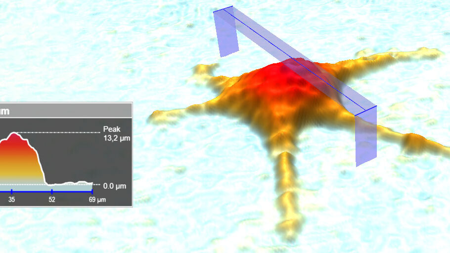
HoloMonitor M4 is a fantastic tool, easy to use, non-invasive, giving very clean 3D cell images. Time-lapse results are fascinating. HoloMonitor carries the researcher in the close vicinity of cells. The software is also very impressive and intuitive, giving unique values on both cell morphology (volume) and behavior (mobility).
DR. ALAIN GELOEN
NATIONAL INSTITUTE OF APPLIED SCIENCES, LYON