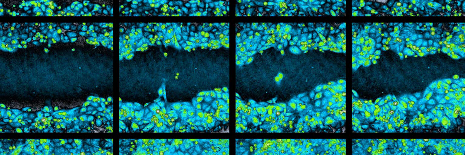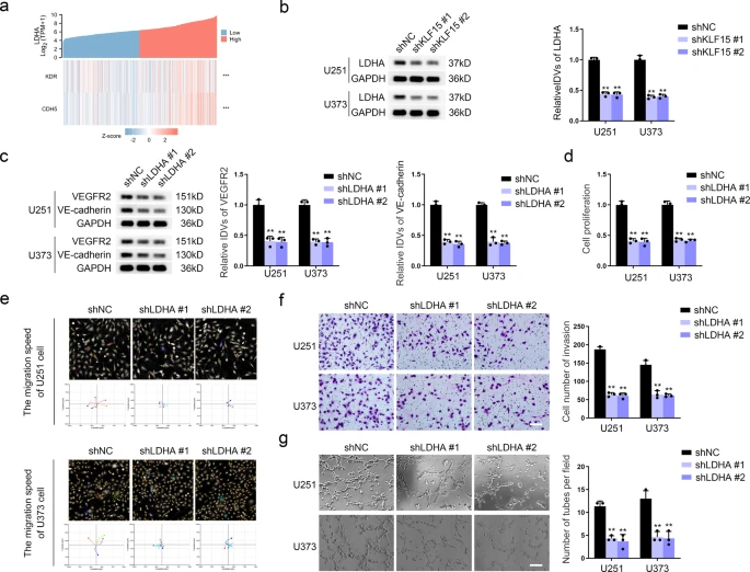
Pseudogene MAPK6P4-encoded functional peptide promotes glioblastoma vasculogenic mimicry development
Journal: Communications Biology (2023)
Research Areas: Cancer research
Cell Lines: U251, U373, and HEK 293T cells
Learn more …

Adipocytes Migration is Altered Through Differentiation
Journal: Microscopy and Microanalysis (2019)
Research Areas: Cell Research
Cell Lines: 3T3-L1
Learn more …

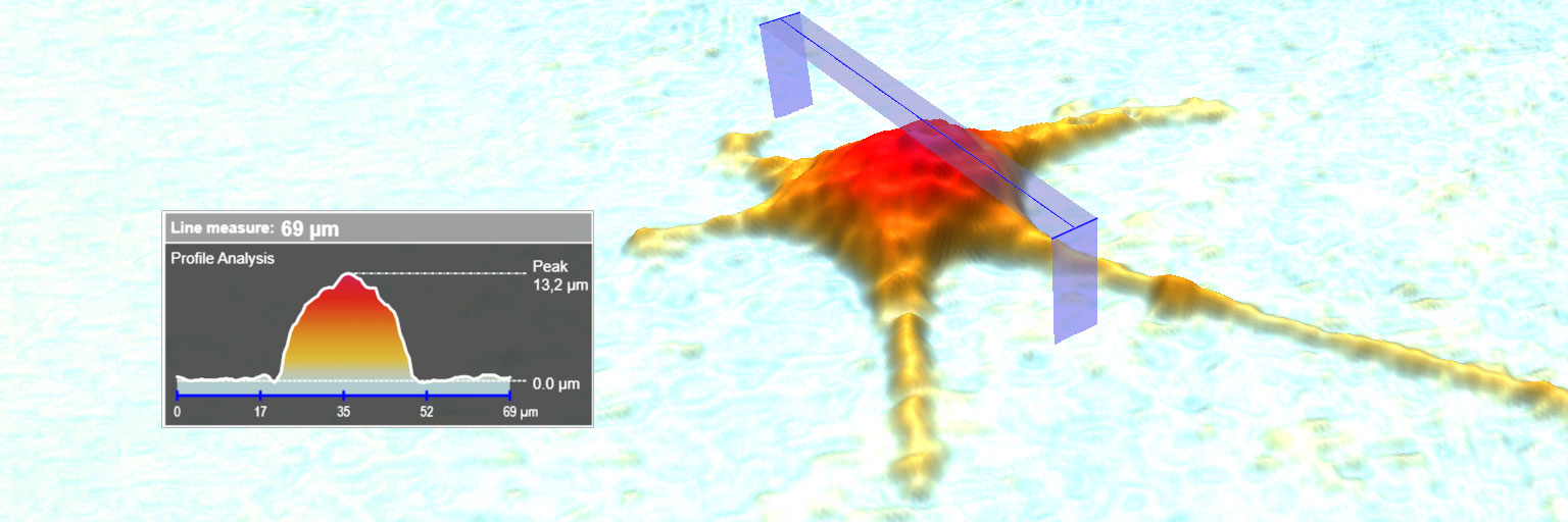
Moving into a New Dimension: Tracking Migrating Cells with Digital Holographic Cytometry in 3D
Journal: Cytometry Part A (2018)
Research Areas: Cancer research
Learn more …

Evaluation of Holographic Imaging Cytometer HoloMonitor M4 Motility Applications
Journal: Cytometry Part A (2018)
Research Areas: Cancer research
Cell Lines: 1205Lu and WM793
Learn more …
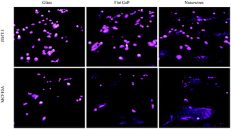
Single cell analysis of proliferation and movement of cancer and normal-like cells on nanowire array substrates
Journal: J. Mater. Chem. B (2018)
Research Areas: Materials of Science
Cell Lines: MCF10A, JIMT-1
Learn more …
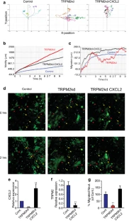
TRPM2 Modulates Neutrophil Attraction to Murine Tumor Cells by Regulating CXCL2 Expression
Journal: Cancer Immunology, Immunotherapy (2018)
Research Areas: Cancer research
Cell Lines: 4Ti, AT3, TRPM2, LLC, human neutrophils
Learn more …