
Prof. Anette Gjörloff Wingren
Faculty of Health and Society, Malmö University
Interviews and News

Best Poster Prize to Louise Sternbaeck
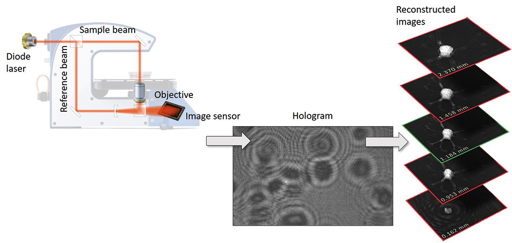
Digital Holographic Microscopy for biomarker detection in cancer

Fighting cancer at an early stage
PHI and Malmö University receives 2.3 million SEK to detect blood-borne cancer cells
EU grants 2.1 million euro to Phase Holographic Imaging and Malmö University with partners for joint cancer research
Researchers propose PHI’s technology to assess reduction in cancer cell growth
Peer Reviewed Articles and Book Chapters

Non-invasive, Label-free Cell Counting and Quantitative Analysis of Adherent Cells Using Digital Holography
Journal: Journal of Microscopy (2008)
Research Areas: Method development
Cell Lines: MCF-10 A, L929, PC-3, DU-145
Digital Holographic Microscopy — Innovative and Non-destructive Analysis of Living Cells
Journal: Microscopy: Science, Technology, Applications and Education (2010)
Research Areas: Method development

Digital Holography and Cell Studies
Journal: Holography-Research and technologies (2011)
Research Areas: Method development
Cells and Holograms — Holograms and Digital Holographic Microscopy as a Tool to Study the Morphology of Living Cells
Journal: Holography — Basic Principles and Contemporary Applications (2013)
Research Areas: Method development

Digital Holographic Microscopy for Non-invasive Monitoring of Cell Cycle Arrest in L929 Cells
Journal: PLOS ONE (2014)
Research Areas: Method development
Cell Lines: L929
Interfacing Antibody-based Microarrays and Digital Holography Enables Label-free Detection for Loss of Cell Volume
Journal: Future science oa (2015)
Research Areas: Cancer research, Drug research
Cell Lines: Jurkat, U2932
Supervised Classification of Etoposide-treated in Vitro Adherent Cells Based on Noninvasive Imaging Morphology
Journal: Journal of Medical Imaging (2017)
Research Areas: Method development
Cell Lines: DU-145
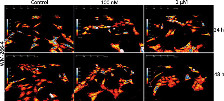
Holography: the Usefulness of Digital Holographic Microscopy for Clinical Diagnostics
Journal: Holographic Materials and Optical Systems (2017)
Research Areas: Method development
Cell Lines: U2932, WM-266-4, CHL-1

Quantitative Phase-contrast Imaging – a Potential Tool for Future Cancer Diagnostics
Journal: Cytometry Part A (2017)
Research Areas: Method development

Moving into a New Dimension: Tracking Migrating Cells with Digital Holographic Cytometry in 3D
Journal: Cytometry Part A (2018)
Research Areas: Cancer research
Digital Holographic Cytometry: Macrophage Uptake of Nanoprobes
Journal: Imaging & Microscopy (2019)
Research Areas: Cytotoxicity
Cell Lines: RAW 264, 7
Evaluation of the Impact of Imprinted Polymer Particles on Morphology and Motility of Breast Cancer Cells by Using Digital Holographic Cytometry
Journal: Applied Sciences (2020)
Research Areas: Cancer research, Materials of Science
Cell Lines: MCF-7 and MDAMB231
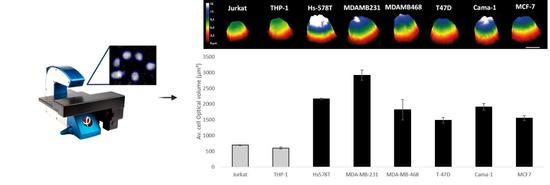
Discrimination between Breast Cancer Cells and White Blood Cells by Non-Invasive Measurements: Implications for a Novel In Vitro-Based Circulating Tumor Cell Model Using Digital Holographic Cytometry
Journal: Applied Sciences (2020)
Research Areas: Cancer research
Cell Lines: Jurkat, THP-1, Hs-578T, MDA-MD-231, MDA-MB-468, T57D, Cama-1, MCF-7