HoloMonitor® Live Cell Imaging System
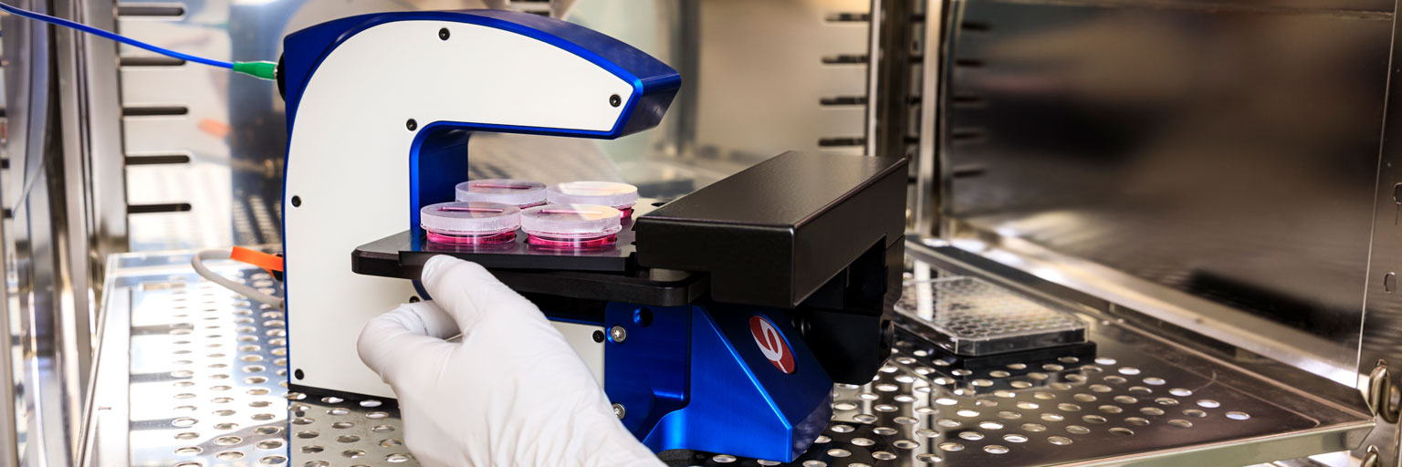
The HoloMonitor® live cell imaging system is your comprehensive single-cell level imaging solution, seamlessly fitting within a standard CO₂ incubator. It includes the compact HoloMonitor live cell microscope and its powerful App Suite software for automated cell imaging and analysis. The fluorescence add-on extends the tool’s versatility and applications even more. We designed HoloMonitor by and for cell biologists, ensuring cell-friendly imaging at its best.
Read more about HoloMonitor in its role as…
A Cell Culture Microscope
The main purpose of the HoloMonitor system is to monitor, record and analyze your cell cultures in the least invasive way possible, even in long-term experiments.
An Incubator Microscope
The compact HoloMonitor microscope is built to work inside your standard incubator. Thus, it ensures stable and physiologically relevant conditions.
A Live Cell Imaging System
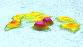
HoloMonitor does not need any labels or stains to visualize your cells. Hence, it has minimal impact on your cells, allowing long-term live cell monitoring.
Why HoloMonitor?
Cell-friendly.
- Avoid known interference of cellular markers for biologically relevant results.
Compact.
- Small footprint to bring imaging inside your incubator.
Affordable.
- Not all live cell imaging microscopes have to be expensive.
Automated.
- Get immediate real-time data and image sequences.
Easy-to-use.
- Intuitive software to guide you from imaging to analysis.
Physiological.
- Controlled conditions matter for your cell culture.

Want to speak to an expert?
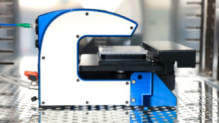
The HoloMonitor Hardware
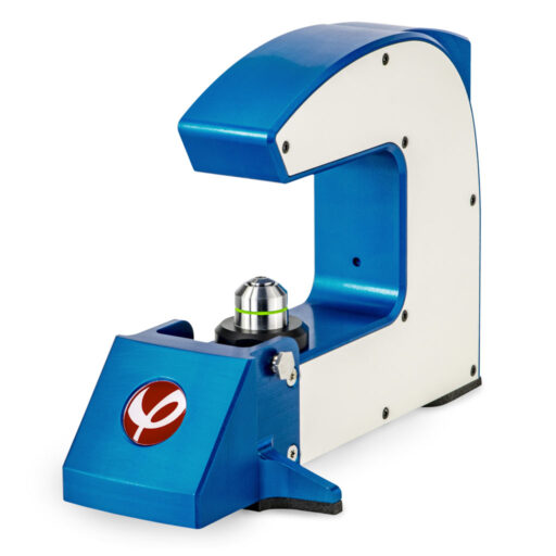
Imaging Unit
The compact base unit is the actual microscope, placed in your incubator together with the motorized stage. It is responsible for continuously imaging your live cell cultures.
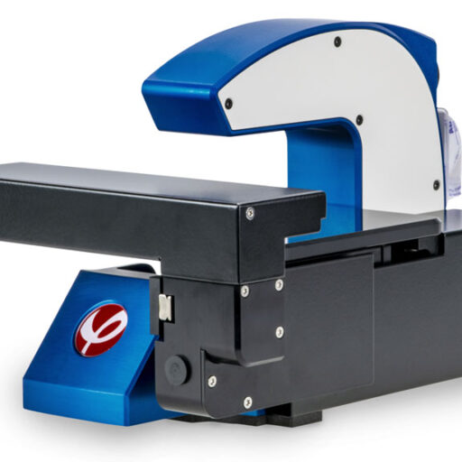
Motorized Stage
The motorized stage supports a variety of culture vessels. Moreover, it enables automated and continuous imaging of multiple positions in your sample.
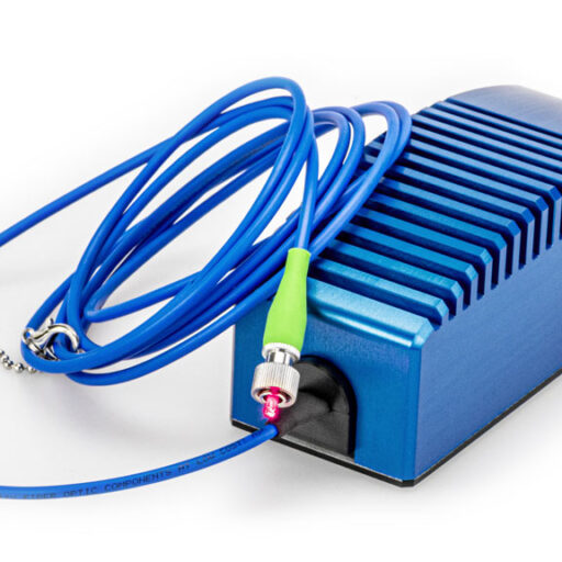
Laser Unit
The laser unit, placed outside your incubator, supplies the system’s low-energy laser for label-free holographic live cell imaging that leaves your cells unaffected.
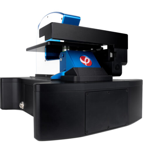
Fluorescence
The add-on fluorescence unit makes it possible to add a green fluorescence dimension to the label-free holography data and study your cells’ behavior in more detail.
Technical Specifications
Technical Information
| Sample stage | Fixed |
| Light source | External laser unit, 635 nm |
| Sample illumination | 635 nm, 0.2 mW/cm² |
| Lateral resolution | 1 µm |
| Field of view | 567 μm × 567μm |
| Working distance | 0.5–2.0 mm |
| Autofocusing range | 1.5 mm |
| Maximum image rate | 1 image/s |
| Image size | 1024 × 1024 pixels |
| Dimensions | 290×200×190mm (W × D × H) |
| Weight | 5.15 kg |
| Cell culture vessels | 6-well, 24-well, 96-well, Petri, IBIDI |
| Power supply | International 110–240 V |
| Interface cable | USB |
| Warranty | 1 year |
Recommended Cell Culture Vessels
Sarstedt 35 mm Petri dishes
Sarstedt 6-well plates
Sarstedt Lumox 24-well plates
Sarstedt Lumox 96-well plates
IBIDI plastic ware (channel slides, chemotaxis slides, wound healing inserts)
We always recommend using our HoloLids™ for optimal optical performance.
Regulatory Compliance
| Electromagnetic compatibility | 2014/30/EU and 2014/35/EU (low voltage directive) |
| Safety requirements | IEC 61010-1:2010 (electrical equipment for measurement, control and laboratory use) |
| Safety of laser products | IEC 60825-1:2014 |
CE marking is subject to the general principles set out in Article 30 of Regulation (EC) No 765/2008.
Requirements
Sample and environment
| Cells | Monolayer of adherent cells |
| Incubator | Access port for cabling |
| Operating temperature | 10–40°C |
| Operating humidity | Max 95% |
Computer
| Operating system | |
| Operating system | Windows 10, 64-bit |
| Processor | Intel Core i7 |
| Memory | 16 GB RAM (8 GB minimum) |
| Hard drive | 512 GB SSD (256 GB minimum) and external USB3 hard drive for backup and transfer |
| Display | Full HD (1920 × 1080) or higher |
For service purposes, it is convenient if the computer has internet access. However, we recommend disabling the internet connection during experiments, as automatic Windows updates may disrupt them.
Technical Information
| Repeatability | 5 µm |
| Travel range | 100 × 70 × 10 mm (X × Y × Z) |
| Dimensions* | 290 × 200 × 185 mm (W × D × H) |
| Operational dimensions* | 375 × 260 × 185 (W × D × H) |
| Minimum space inside the incubator | 400 × 270 × 185 (W × D × H) |
| Weight* | 5.5 kg |
| Operating temperature | 10–40°C |
| Operating humidity | Max 95% |
| Cell culture vessel support | 6-well, 24-well, 96-well, Petri, IBIDI |
| Warranty | 1 year |
*Including the base unit
Technical Information
| Exposure time | 5 ms, non-scanning |
| Light source | External laser unit, 635 nm |
| Sample illumination | 0.2 mW/cm² |
| Power supply | International 110–240 V |
| Interface cable | USB |
| Warranty | 1 year |
Technical Information
| 1-channel fluorescence | Optimized for EGFP (FITC-Cy2) |
| Excitation | 470 nm filtered LED, with bandwidth (FWHM): 40 nm |
| Emission | 525 nm filtered, with bandwidth (FWHM): 50 nm |
| Exposure time | 1 – 1000 ms |
| Gain | 1 – 4 |
| Magnification | 10x, matched with mounted HoloMonitor M4 |
| Sensor | 1280 x 1024 px, 1.31 MPix |
| Image Resolution | 0.5 µm |
| Field of view | 567 μm × 567μm |
| Image size | 1024 × 1024 pixels |
| Dimensions | 310 x 180 x 85 mm (W × D × H) |
| Weight | 4.0 kg |
| Power | < 5 W |
| Interface | USB connector |
| Software requirement | HoloMonitor App Suite software 4.0 |
| Operating environment | 10 – 50 °C, < 95 % relative humidity |
The HoloMonitor Software
The heart of HoloMonitor is the modular App Suite software, with tailor-made apps for different live cell assays. Each app offers a guided setup of your experiment, automatic image collection and real-time data analysis and presentation.
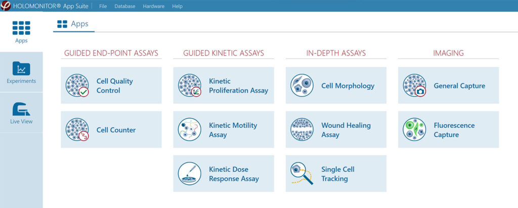
Moreover, App Suite excels when it comes to data re-analysis. Every experiment contains all the data necessary to reanalyze it as another type of experiment. For example, a proliferation assay can be reanalyzed as a motility assay at a later point. Hence, no need to spend time, money and cells on a new experiment!
HoloMonitor Live Cell Assays
The Workflow
Designed with cell biologists, the HoloMonitor App Suite software offers a simple and intuitive user interface, guiding you through the workflow from experiment setup to data presentation in 5 steps.
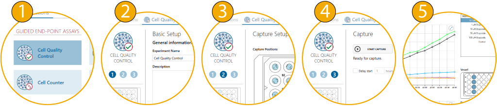
In addition, you can easily create colorful videos and images to present your results. Of course, all data can also be exported to Microsoft Excel for further analysis.
Best microscope I’ve tried so far—worth every penny!
Markus Bosteen
Senior Scientist, Novo Nordisk, Copenhagen
HoloMonitor Accessories
Culture Vessel Holders
HoloMonitor’s motorized stage supports a variety of culture vessels, each secured with a vessel holder. This means easy attachment and exact positioning on the microscope.
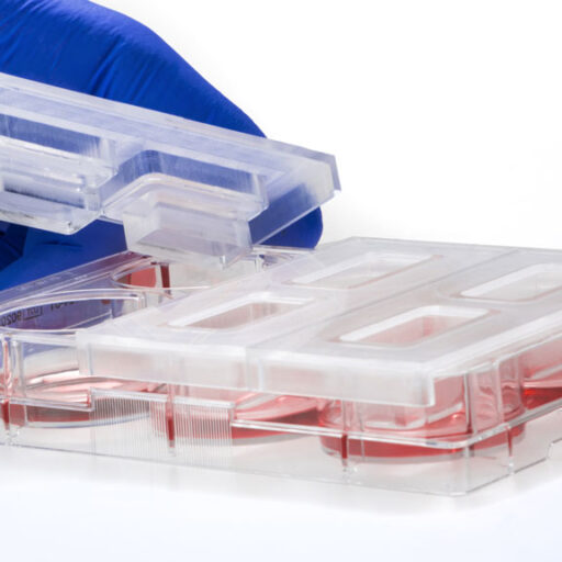
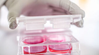
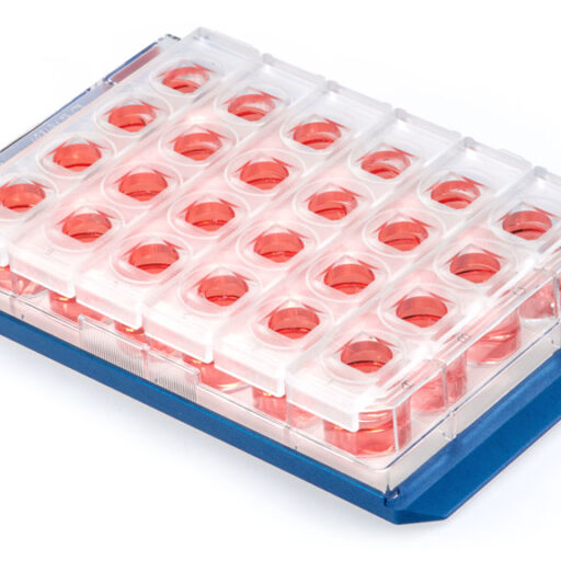
HoloLids™
HoloLids are specially designed lids for standard cell culture vessels. They are optically superior to normal vessel lids and ensure the best possible image quality every time.
HoloDry™
HoloDry is a non-toxic canister that is easily attached to the HoloMonitor microscope. It keeps the optics and interior free of condensation, which ensures optimal performance.