A Compact Live Cell Imaging System For Your Incubator
Compact
to fit in your standard incubator

Non-invasive
for more biologically relevant results

Accurate
with exact cell segmentation

Meet HoloMonitor®
Our Live Cell Imaging system
- Measure 30+ cellular features from population down to single cell level
- Non-invasive imaging, directly in your standard incubator
- Easy and comprehensive data presentation in real time
- Easy-to-use, guided software that lets you reanalyse your experiments
Advanced Live Cell Imaging with HoloMonitor
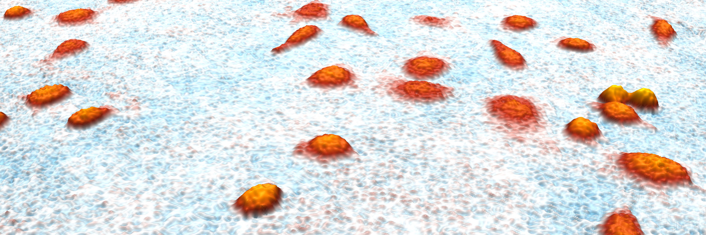
Live cell imaging is where the HoloMonitor microscope excels. Using digital holography, it collects 3D images of your cell cultures completely non-invasive. It requires no labels or stains, and the low-energy laser generates negligible phototoxicity, therefore, you can reuse your cells for downstream experiments. Placed in your standard incubator, this allows you to collect real-time data on your cell’s unaffected behavior, as often and as long as you want.
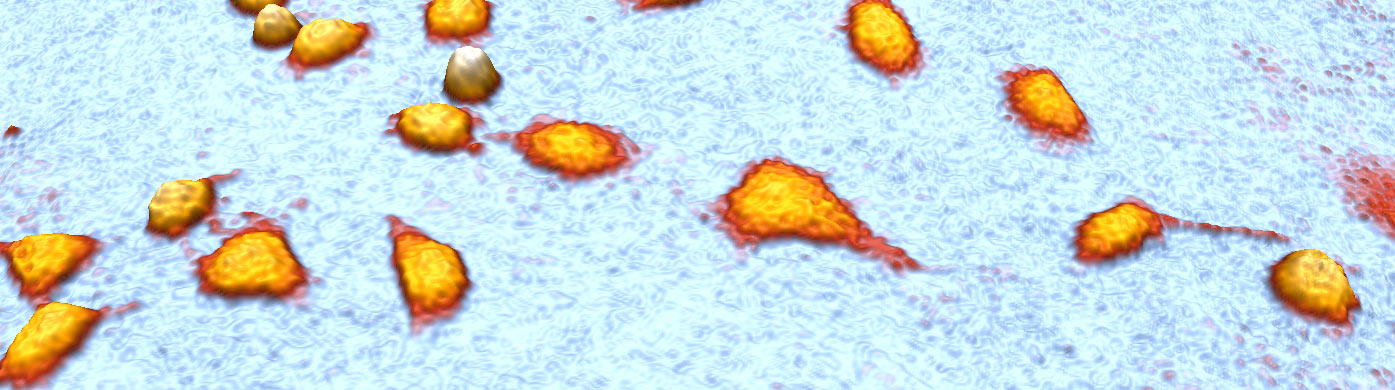
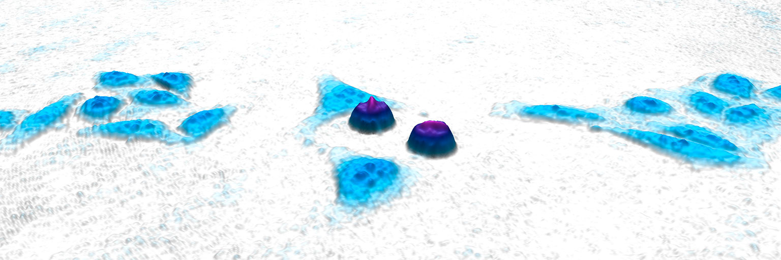
This time-lapse video was recorded with HoloMonitor over 24 hours. It shows MDA-MB-231 breast cancer cells in vitro. The images are captured completely label-free and the colors are added afterward in the computer.
Your 8-in-1 Cell Analysis Tool

Quantifying over time is crucial for a full understanding of cell systems. I am convinced that time-lapse cytometry will enable the next level of insight.
PROF. TIMM SCHROEDER
ETH ZÜRICH
The Applications
Kinetic Cell Morphology
(with 30+ parameters)
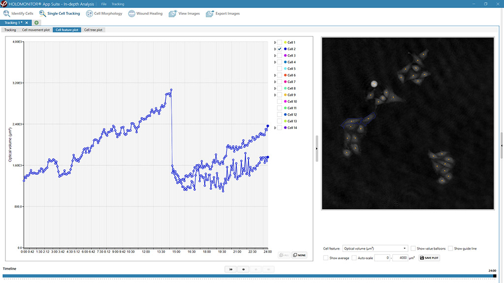
In-depth Single Cell
Tracking & Analysis
Kinetic Cell Motility &
Migration
Wound Healing
(Scratch assay)
Chemotaxis
Kinetic Cell Proliferation
Cell Growth | Cell Division
Cell Cycle
Cellular Differentiation
Kinetic
Drug Dose-Response
Cytotoxicity & Cell Death
Cell Counter
Cell Quality Control (QC)
Why HoloMonitor?
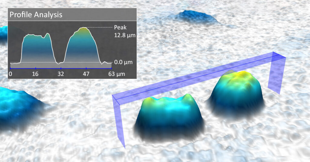
Always measures over 30 cellular features
Every image HoloMonitor takes contains position and size information about every cell, in three dimensions. From this, you can extract over 30 different cellular features, such as cell area, thickness, volume, and migration speed. Moreover, many of the features are unique for digital holography microscopy. With a live cell imaging time-lapse, you can study all of these parameters simultaneously over time, at population level all the way down to single-cell level.
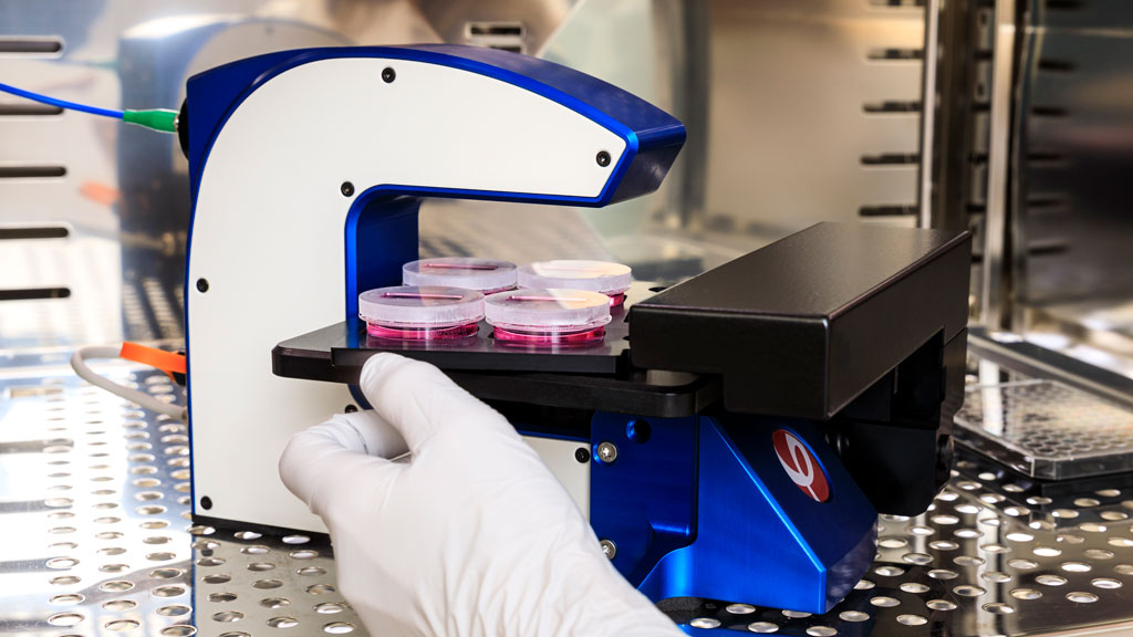
Non-invasive incubator-based cell imager
The HoloMonitor live cell imaging system is designed to be placed inside your standard incubator, lab bench, or hypoxia chamber 24/7, so allowing you to image your cells in their natural environment. Furthermore, HoloMonitor uses digital holography to image cells without any labels or stains, reducing the imaging influence on cell behavior to a minimum. In summary, this makes HoloMonitor the perfect, non-invasive live cell imaging tool, unrivaled by the competition.
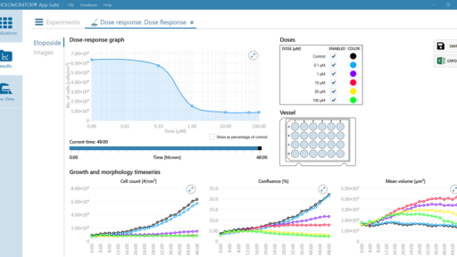
Clear and comprehensive data presentation
The HoloMonitor App Suite live cell imaging software collects all data on your cells and presents it the way you want. For example, you can get graphs showing cellular changes over time, dot plots to find subpopulations within your cell cultures, or just plain numbers for the cell count. Of course, you can easily export all data can be to Microsoft Excel as well, allowing you to do your own calculations and data presentation.
Strong software with walk-through assays
The HoloMonitor App Suite live cell imaging software offers several predefined live cell assays to study, for example, proliferation and morphology. The software guides you through an easy setup, runs the experiment for you, and compiles the results in real-time. Moreover, you can re-analyze your results with other HoloMonitor assays to generate more data without setting up new experiments, to save your time, money, and cells.
Novel Live Cell Imaging System
Wound healing (scratch assay) with HoloMonitor. The software automatically gives you relevant data such as cell front velocity and gap closure over time.
Single cell tracking with HoloMonitor. Every cell is automatically tracked and the software generates cell trees and motility data on the fly.