
Meet HoloMonitor® NG
The next-generation QPI platform for live cell imaging
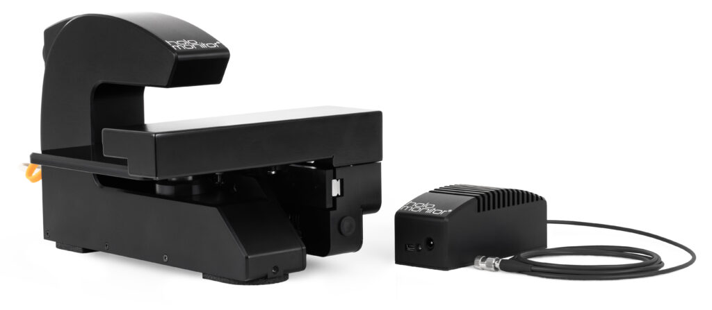
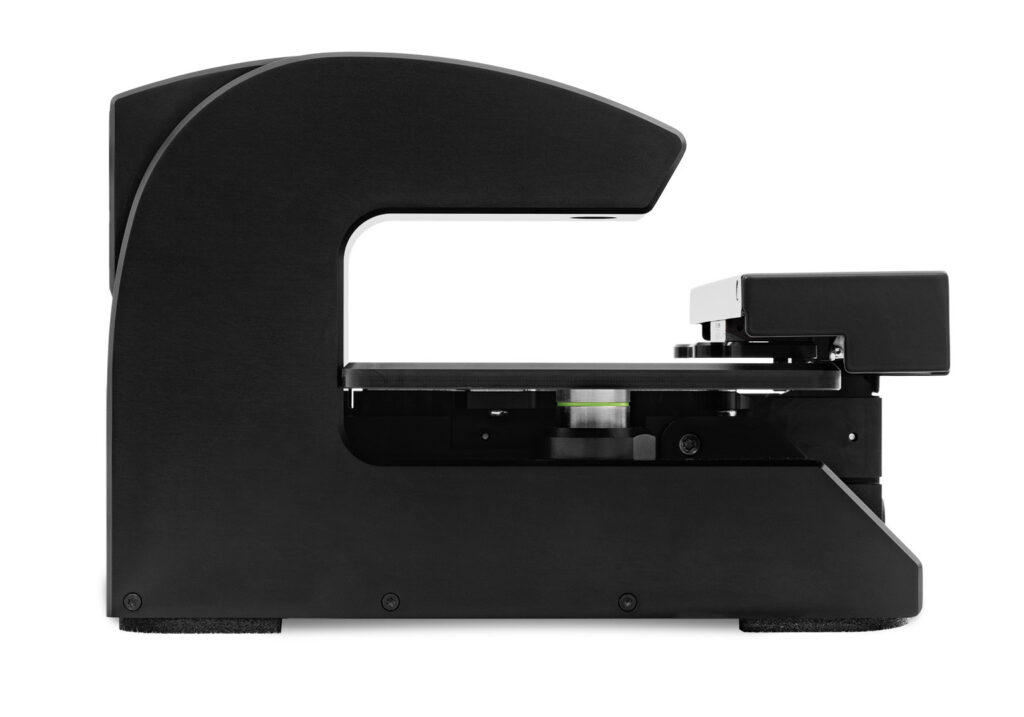
Phase Holographic Imaging introduces HoloMonitor® NG—our next-generation, adaptable QPI platform for live cell imaging. With a redesigned hardware architecture and significantly improved software, we deliver an instrument with fourfold higher resolution and more accurate, automated segmentation. Built with clinical workflows in mind, HoloMonitor NG is engineered to meet the rigorous requirements set by clinical settings.
Major Highlights
Detailed images of thin cells
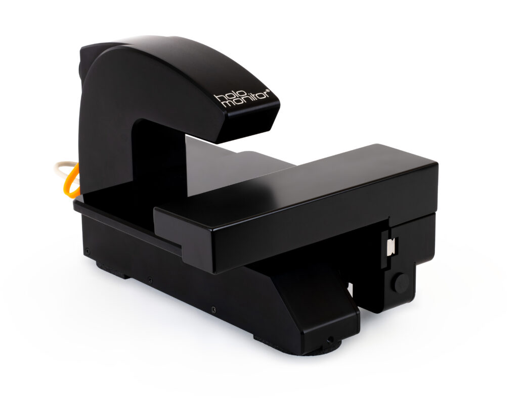
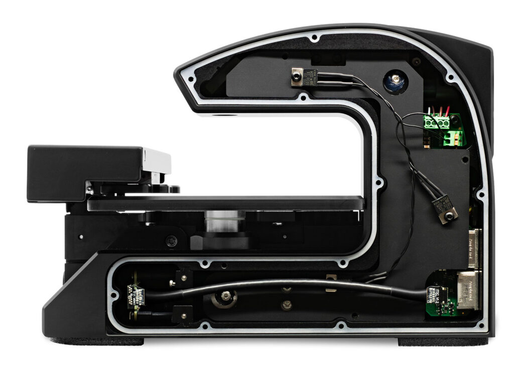
Our hardware engineers have reworked the internals of HoloMonitor NG, with an all-new camera at the core. With higher resolution and significantly reduced background noise, it allows for finer detail and improved imaging of thin cells.
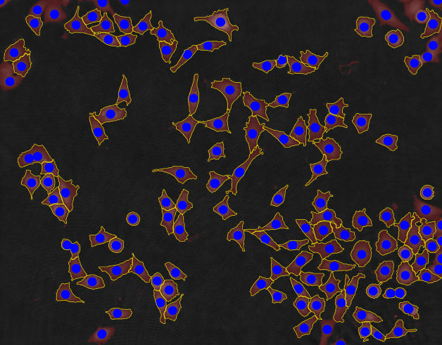
Precise cell identification and analysis
Thanks to the improved hologram quality, our software team has been able to significantly enhance and automate the segmentation algorithm. It now requires minimal user input to deliver accurate cell segmentation.
Sterile inside and out
To justify its position in a clinical setting, the new HoloMonitor NG can be thoroughly sterilized using ethylene oxide. It is a powerful and commonly used sterilant in life science, ensuring a sterile environment for sensitive cells.
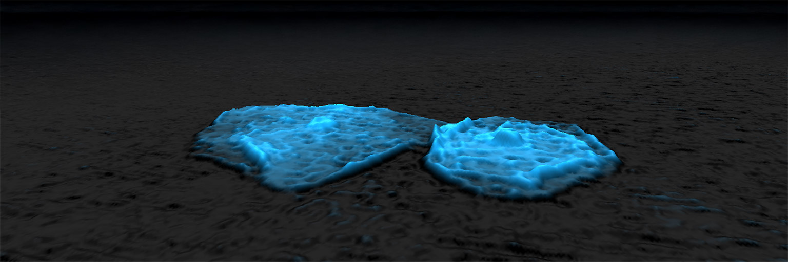
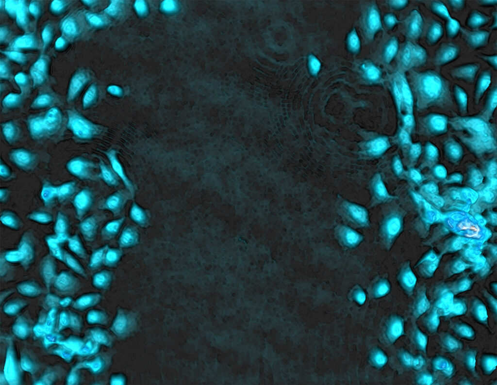
Primary epithelial cells—about 2 µm thick
Redesigned hardware improves image quality for thin cells
HoloMonitor NG has been equipped with a brand new camera sensor, modified specifically for this instrument, along with a redesigned PCB engineered to meet the substantially higher data throughput. The camera delivers 4 megapixel holograms with minimal noise, resulting in quantitative phase images with unprecedented quality and more reliable segmentation and analysis. The improved resolution and signal quality reveal finer cellular detail, but more importantly, it also makes it easier to image thinner cells—a characteristic commonly seen in primary cells and clinical samples—expanding the range of applications where label-free holographic imaging can be used with confidence. Furthermore, the new system is designed for flexibility, making it well suited for the specific demands of applications such as IVF, MSC research, and cardiomyocyte studies—areas that often require higher frame rates or a larger field of view. Its adaptable architecture makes it possible to tailor the platform as requirements evolve.
Do you want to know more?
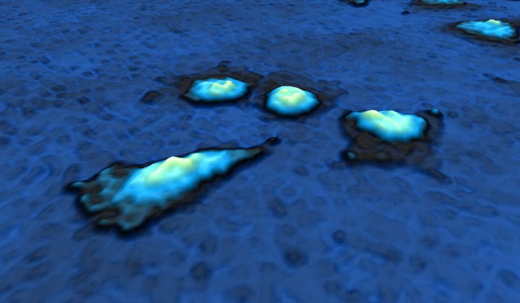
Enhanced segmentation for reliable data
With cleaner, higher-resolution holograms and reduced noise, our software team has been able to develop an updated reconstruction algorithm that produces more reliable quantitative phase images and a more robust assessment of image quality. In parallel, the stronger signal and increased detail in the reconstructed images have enabled major advances in segmentation.
The new segmentation algorithms are more accurate and consistent across varying cell morphologies, and provides automated cell segmentation for reduced manual intervention. This makes it easier to set up assays and obtain reproducible single-cell measurements at scale—especially in challenging samples where thin or low-contrast cells previously limited performance.
HoloMonitor NG is prepared for AI-enabled workflows, allowing users to train AI-driven segmentation models on their own unique cell types. This enables even greater automation and more reliable, consistent segmentation across experiments.
Meet HoloMonitor NG and the new App Suite 5.0 software
Sterilization for a safe cell environment
HoloMonitor NG is designed to support clinical workflows and can be thoroughly sterilized using ethylene oxide (EtO). EtO is a powerful and well-established sterilant for heat sensitive medical devices and is widely used in life science applications where sterile handling is essential. Aseptic practice is critical for obtaining reproducible, biologically relevant results, and EtO sterilization enables the HoloMonitor NG hardware to remain sterile throughout contact with sensitive, biological samples.

HoloMonitor based quantitative phase imaging enables label-free analysis of fundamental cellular behaviors central to asthma pathology. By capturing mast cell–driven effects on cell migration, morphology, and epithelial repair, this approach provides mechanistic insight into impaired wound healing and airway remodeling—key features of asthmatic disease.
Cecilia Andersson, Docent
Lund University
Pre-clinical and clinical studies
HoloMonitor NG has been used in an ongoing clinical study of uncontrolled asthma to track wound closure, morphology, and migration in patient-derived bronchial epithelial cells—continuously and label-free using quantitative phase imaging. Early exploratory observations highlight the potential of holographic live-cell imaging as a functional complement to established clinical biomarkers.
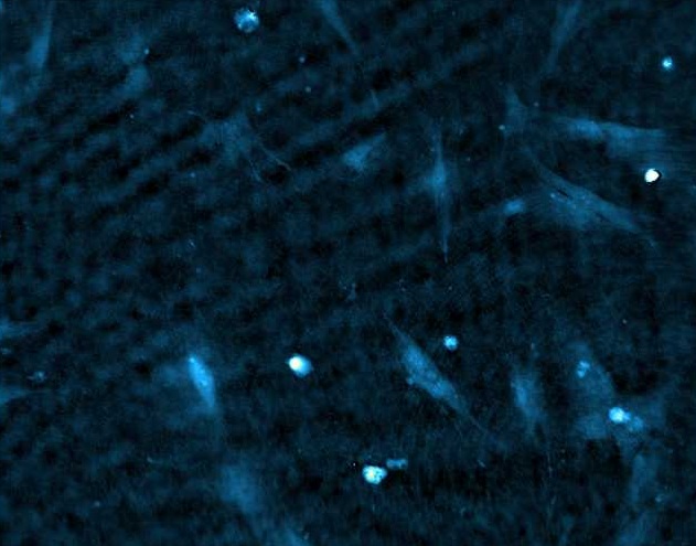
Together with a partner at Wake Forest Institute of Regenerative Medicine (WFIRM) in Winston Salem, NC, USA, we study human primary placental cells. These very thin cells have been challenging but going forward, HoloMonitor NG will facilitate imaging and analysis.
Do you want to know more?