Recommended Cell Culture Vessels
To ensure optimal image quality when using HoloMonitor®, we have compiled a list of thoroughly tested cell culture vessels that do not interfere with the holographic image. As optically low-quality plastics can compromise the image quality and hence the analysis, using the tested vessels listed below is highly recommended. For superior image quality, HoloLids™ should be used to further eliminate image disturbances.
| Cell culture vessel | Manufacturer | Type | Catalogue no. | Compatible with HoloLids™? |
|---|---|---|---|---|
| ibidi µ-Dish, 35 mm, high | ibidi | Culture dish | 81156 | Yes |
| ibidi µ-Dish, 35 mm, high, with Culture-Insert, 2 well | ibidi | Culture dish | 81176 | Yes* |
| ibidi µ-Slide Chemotaxis | ibidi | Slide | 80326 | Not applicable |
| ibidi µ-Slide I | ibidi | Slide | 80106 | Not applicable |
| ibidi µ-Plate, 24 well | ibidi | 24 well plate | 82426 | Yes |
| ibidi µ-Plate, 24 well, with Culture-Insert, 2 well | ibidi | 24 well plate | 80242 | Yes* |
| Sarstedt lumox® multiwell, 24 well | Sarstedt | 24 well plate | 94.6000.014 | Yes |
| Sarstedt lumox® multiwell, 96 well | Sarstedt | 96 well plate | 94.6000.024 | Yes |
| Sarstedt Tissue culture plate, Standard, 6 well | Sarstedt | 6 well plate | 83.3920 | Yes |
| Sarstedt Tissue culture dish, 35 mm | Sarstedt | Culture dish | 83.3900 | Yes |
| Countess™ Cell Counting Chamber Slide | Invitrogen | Slide | C10228 | Not applicable |
*HoloLids™ can be used after the Culture-Insert has been removed.
Please contact our team if you can’t find your preferred cell culture vessel in the list or have any questions regarding vessel compatibility with HoloMonitor.
Why do we recommend these particular vessels?
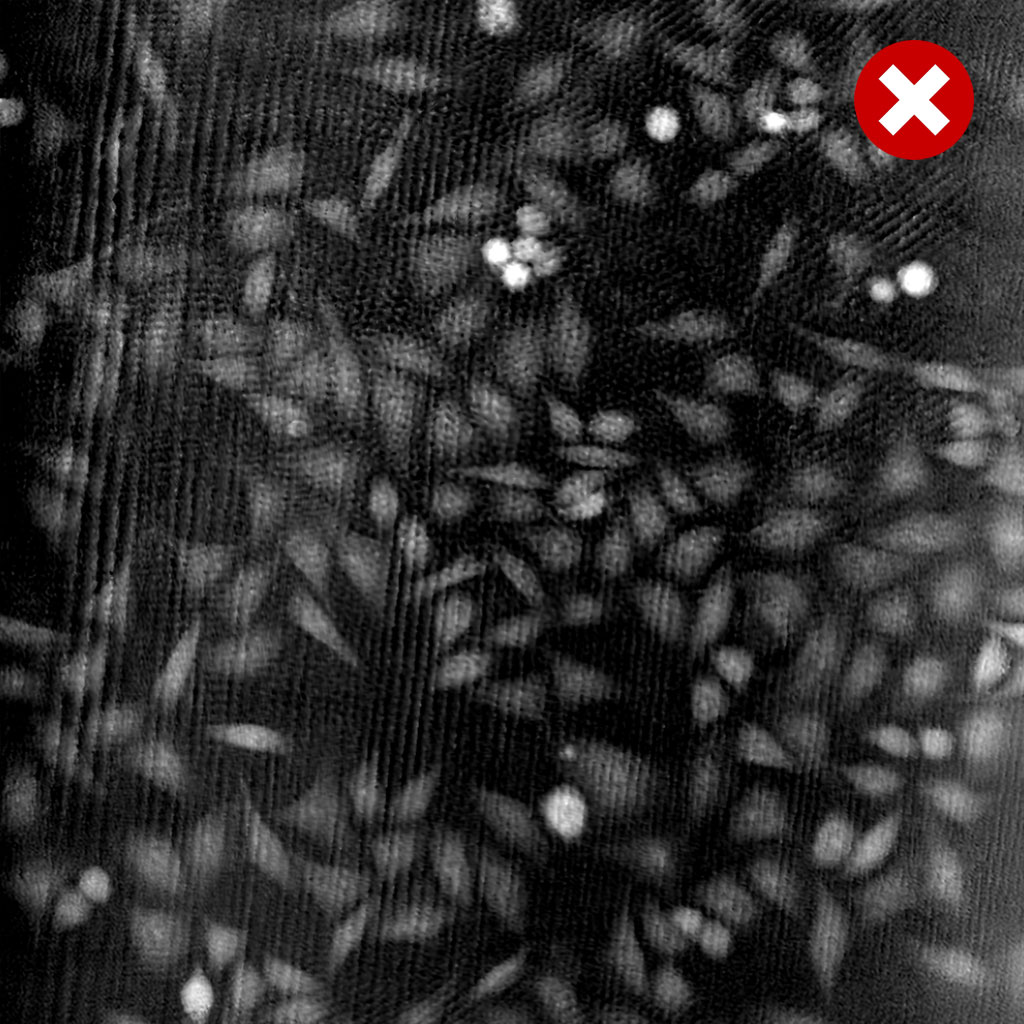
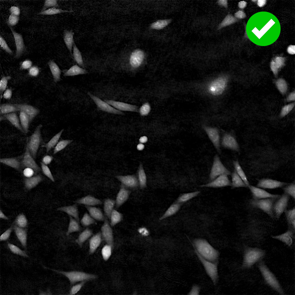
HoloMonitor uses Quantitative Phase Imaging (QPI) to image living cells. This technology quantifies the phase shift that occurs as light passes through the cells, enabling precise calculations of the cell morphology in three dimensions. It is important, however, that the phase shift is only induced by the cells and remains unaltered as it passes through the plastic of the cell culture vessel. Any plastic impurities, inhomogeneities, or variations in refractive index can distort the light and result in less accurate cell measurements, introduce noise into the image and ultimately result in less precise segmentation and morphology calculations. Below are two QPI images, one captured on optically low-quality plastic and the other on optically high-quality plastic, illustrating the impact of plastic quality on the resulting image. The vessels listed above have been thoroughly tested by us and have shown consistently high optical quality.