Customer Publication
Quantifying the Rate, Degree, and Heterogeneity of Morphological Change during an Epithelial to Mesenchymal Transition Using Digital Holographic Cytometry
Journal: Applied Sciences (2020)
Institution: Lund University
Research Areas: Cancer research
Cell Lines: HDF, RPTE, L929, NMuMG, JIMT-1 (HDF – human derman fibroblasts; RTPE – human renal proximal tubule epothelial cells; L929 – mouse fibroblast cell line; NMuMG – mouse mmary gland cells; JIMT-1 – human breast cancer cell line)
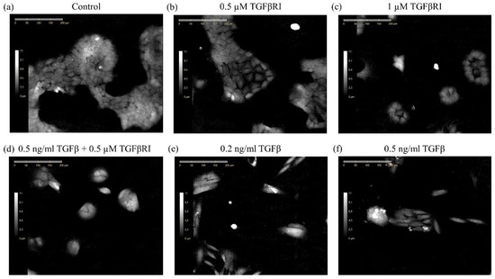
Summary: Cell-based assays designed to characterize epithelial-mesenchymal phenotypic (EMP) transition usually are end-point, bulk population, or qualitative methods. Thus obtained results can miss key information on precise EMP transition process nature, kinetics, and uniformity of molecular changes depending on cell line and conditions. In this work, S. Kamlund with colleagues used digital holographic cytometry (DHC), HoloMonitor M4, to develop a tractable method for monitoring EMP in adherent mammalian cell populations. Authors have shown, that cells in different EMP transition stages exhibit different cell morphological parameters (e.g. cell eccentricity, cell hull convexity, cell optical thickness, cell roughness skewness, etc), which can be quantified and applied to calculate the degree of the EMP transition. This could potentially make DHC and HoloMonitor M4 a fast and efficient primary method to investigate EMP cell morphological changes due to experimental conditions.