Chapter 6
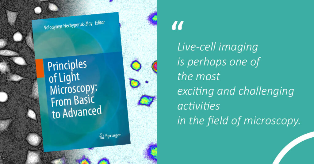
Live-Cell Imaging: A Balancing Act Between Speed, Sensitivity, and Resolution
Principles of Light Microscopy: From Basic to Advanced | January 29, 2023
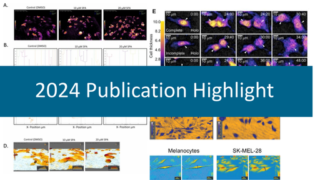
How is label-free digital holography a gentle cell imaging method?
Thank you to the authors for including and highlighting our HoloMonitor® technology in this chapter as a gentle time-lapse imaging method for non-invasive, long-term and quantitative analysis of living cells.
About:
What are the technological developments in the rapidly evolving field of live-cell microscopy?
Live-cell imaging is perhaps one of the most exciting and challenging activities in the field of microscopy.
It is exciting as recent developments in microscope technology have enabled scientists to visualize cellular and subcellular processes in real-time down to the molecular level. With this comes the prospect of studying the mechanisms of diseases in greater detail and finding possible therapeutic solutions.
Nevertheless, live-cell imaging is equally challenging because cells themselves and all cellular processes are extremely sensitive to the impact of using light for their visualization.
This chapter aims to provide a practical overview for early Ph.D. students and more experienced post-docs, who will spend considerable time mastering the most important challenges and prerequisites in the very rapidly evolving field of live-cell microscopy.
If you are a researcher and would like to learn more about digital holography applications for your type of research and cells, you’re always welcome to contact our team!