HoloMonitor® Live Cell Videos & Images
Below, a selection of representative live cell videos and images are presented from various experiments using HoloMonitor — a holographic live cell imager.
Researchers across the world employ HoloMonitor to automatically obtain live cell morphology, proliferation and migration data on a single-cell level, in real-time. HoloMonitor operates from within a standard cell incubator or hypoxia chamber to non-invasively provide cell-friendly cell analysis over extended periods.
Video Galleries
![Our Story Banner](https://phiab.com/wp-content/uploads/2020/08/Our-story-banner-320x180.jpg)
The PHI Story
Get to know PHI! PHI provides innovative tools that let you continuously image and monitor your cells directly in your incubator.
Learn more …
![The HoloMonitor M4 cell culture microscope on a lab bench next to cell culture accessories](https://phiab.com/wp-content/uploads/2021/03/phi-2-20210210_3047hu-320x180.jpg)
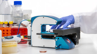
HoloMonitor How-to Tutorials
Learn how to handle the HoloMonitor Live Cell Imaging & Analysis System.
Learn more …
![Closeup of the HoloMonitor live cell imaging microscope on a lab bench.](https://phiab.com/wp-content/uploads/2019/06/T17A3552-320x180.jpg)
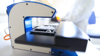
Introducing HoloMonitor M4
Get to know HoloMonitor M4 — an innovative, label-free live cell imaging and analysis system and its software App Suite!
Learn more …
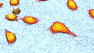
![Cell Banner](https://phiab.com/wp-content/uploads/2020/01/cells-320x180.jpg)
Live Cell Imaging Videos
Live cell videos, imaged without labels, directly in an incubator by the HoloMonitor imaging system.
Learn more …
Never miss the latest news on label-free live cell imaging!
Follow us on social media!
Image Gallery
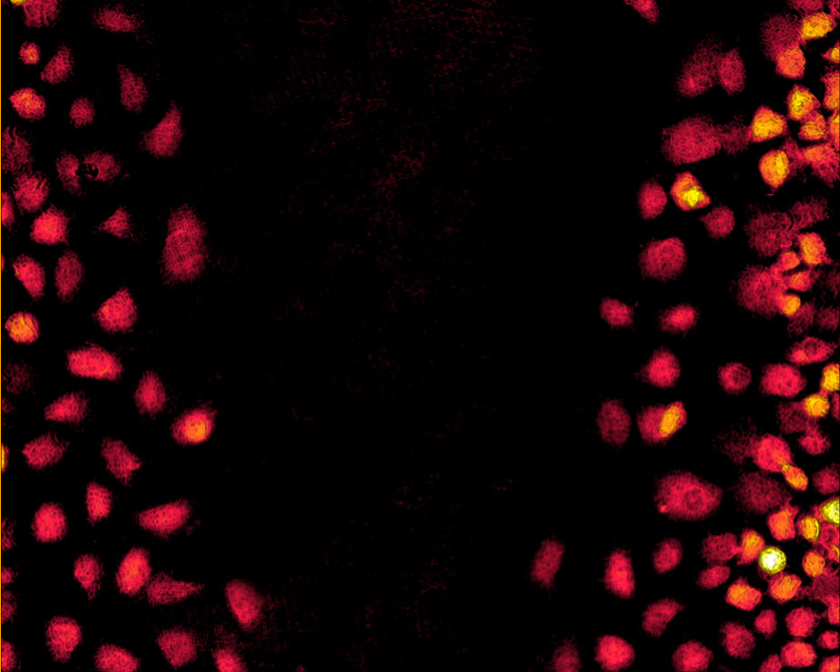
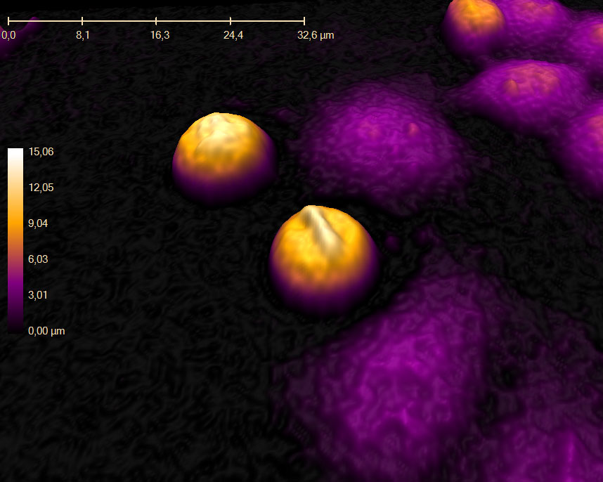
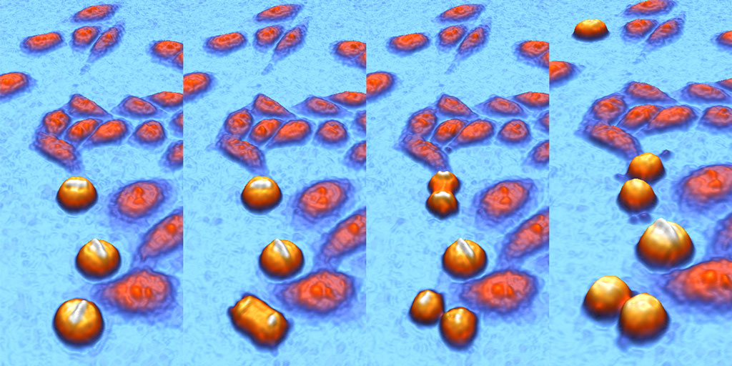
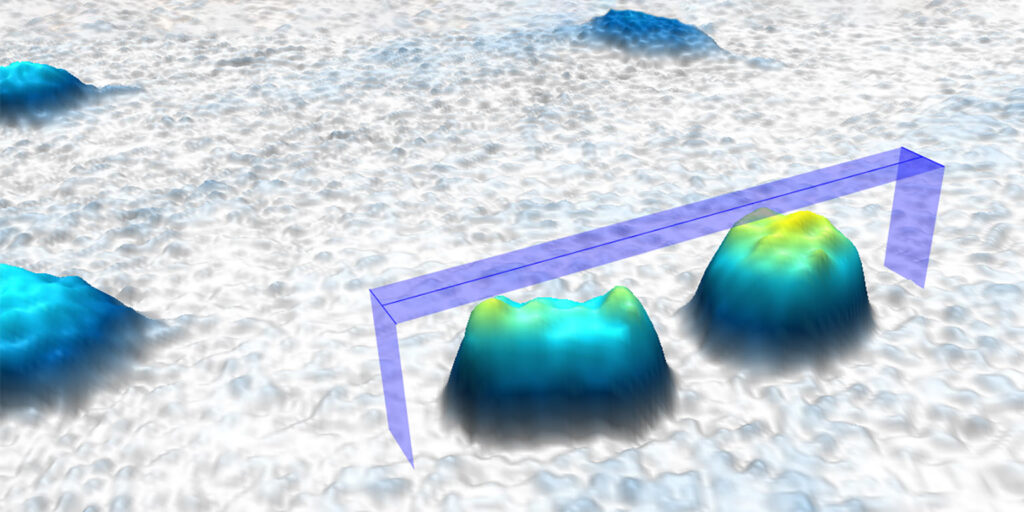
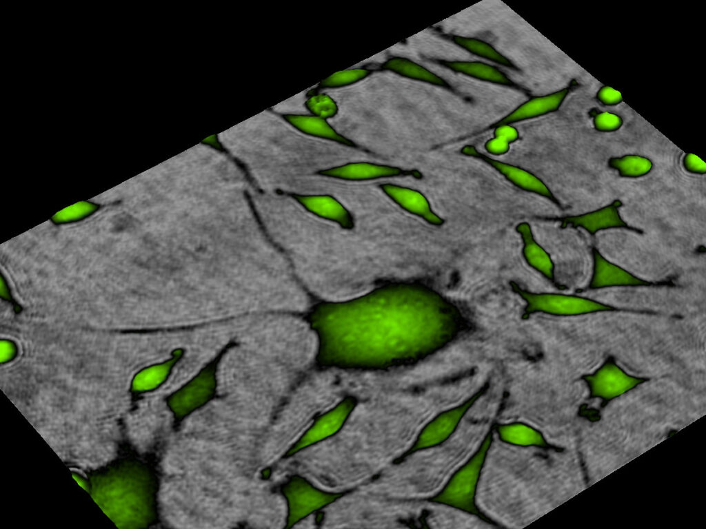
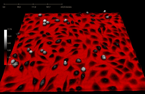
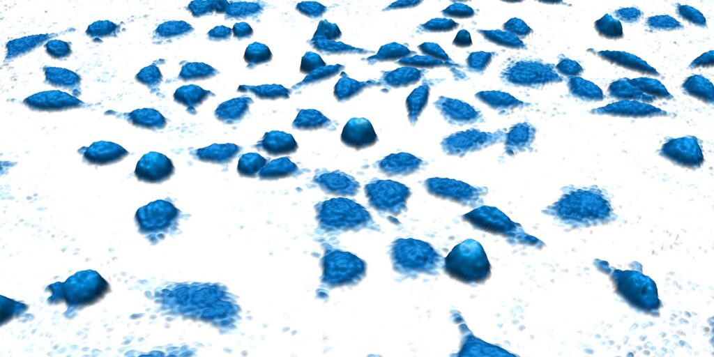
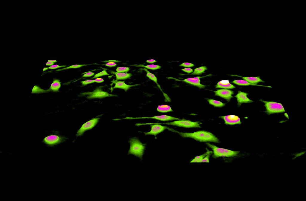
All HoloMonitor applications are label-free. The cell colors below are added by the HoloMonitor cell imaging software — App Suite.
![Jimt-1 cells](https://phiab.com/wp-content/uploads/2020/08/Jimt-cells-in-3D-banner-1024x341.jpg)
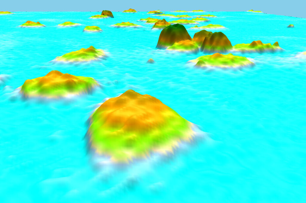
![DU-145 cells imaged from an angle by the HoloMonitor live cell imager](https://phiab.com/wp-content/uploads/2017/10/CellAtoll-1024x680.jpg)
![Digitally colored L929 cells imaged by HoloMonitor](https://phiab.com/wp-content/uploads/2019/08/L929-Candy-banner-1024x341.jpg)
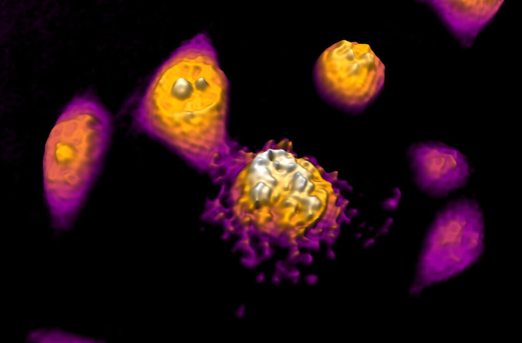
![Apoptotic DU-145 Cell imaged using the HoloMonitor cell culture microscope](https://phiab.com/wp-content/uploads/2017/10/Apoptotic-DU-145-1024x673.jpg)
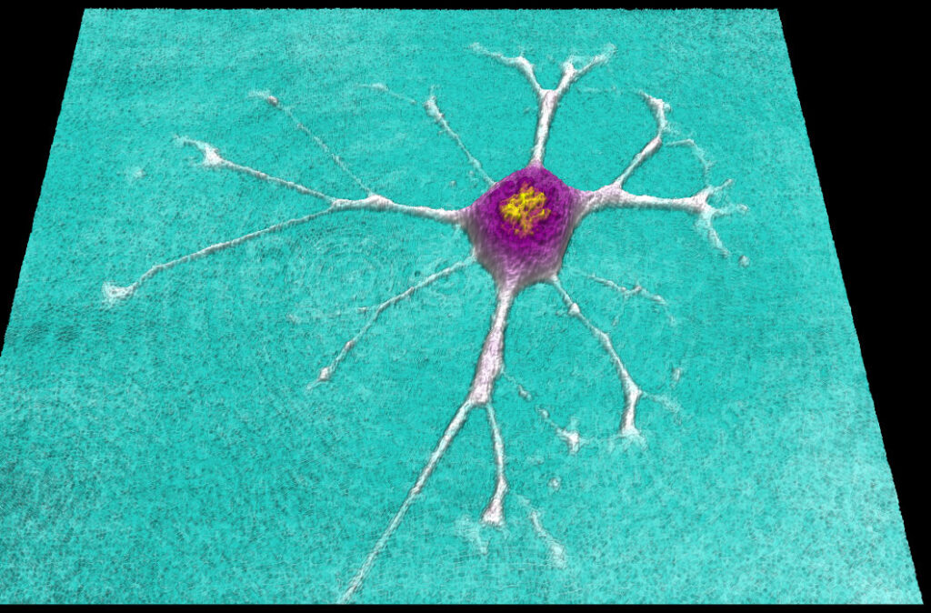
![Differentiated 3T3 cell imaged using the HoloMonitor live cell imaging system](https://phiab.com/wp-content/uploads/2017/10/Differentiated-3T3-1024x674.jpg)
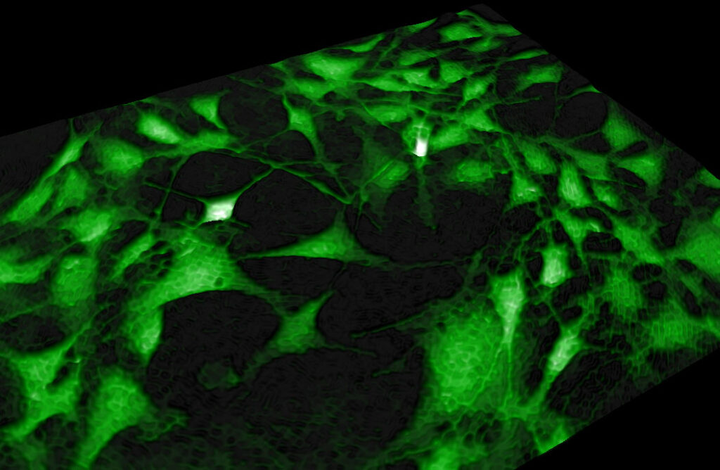
![Green cells on 3D surface, imaged by the HoloMonitor live cell imager](https://phiab.com/wp-content/uploads/2018/03/MEF-in-green-1024x668.jpg)
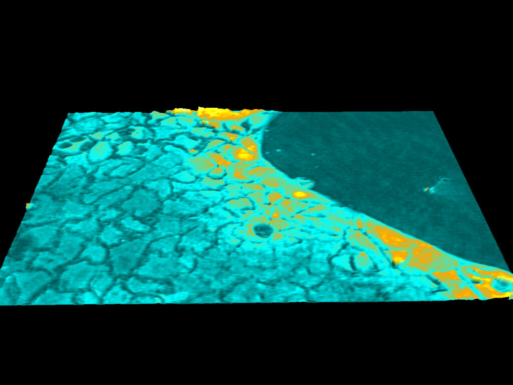
![Wound healing assay of Jimt-1 cells, imaged by the HoloMonitor live cell imaging system](https://phiab.com/wp-content/uploads/2017/10/Wound-healing-Jimt-1.jpg)
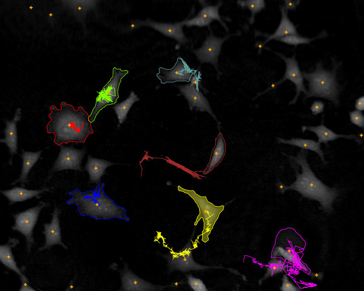
![Cells tracked with the HoloMonitor cell tracking software.](https://phiab.com/wp-content/uploads/2017/10/Cell-motility-L929.jpg)
![Mitosis of Pan-C cells, created by the HoloMonitor cell culture imager and microscope](https://phiab.com/wp-content/uploads/2018/03/Mitosis-Pan-C.jpg)
![Sheet of CaCo-2 cells imaged by the HoloMonitor live cell imaging microscope](https://phiab.com/wp-content/uploads/2021/01/CaCo-2-1024x768.jpg)
![Sequence of images showing cell divisions, created by the incubator-based cell culture microscope - HoloMonitor](https://phiab.com/wp-content/uploads/2020/11/cells-banner-relevant-events.jpg)
![Cell thickness measurement](https://phiab.com/wp-content/uploads/2020/09/cells_bar_1536x768-1024x512.jpg)
![Unstained partially colored](https://phiab.com/wp-content/uploads/2020/08/HoloMonitor-cell-banner-1024x341.jpg)
![Huge L929 in green imaged and colored by the HoloMonitor software in green](https://phiab.com/wp-content/uploads/2021/01/Huge-L929-in-green-1024x768.jpg)
![Proliferating L929 cells](https://phiab.com/wp-content/uploads/2017/12/Cell-death-untreated-L929-cells-1.png)
![](https://phiab.com/wp-content/uploads/2020/09/live-cell-imaging-system-lp-1024x512.jpg)
![](https://phiab.com/wp-content/uploads/2020/02/banner-1024x341.jpg)
![](https://phiab.com/wp-content/uploads/2020/05/online-demo-1024x341.jpg)
![](https://phiab.com/wp-content/uploads/2018/02/banner-cell_Movement-1024x341.jpg)
![Green colored cells, imaged by the HoloMonitor cell culture microscope](https://phiab.com/wp-content/uploads/2017/10/Green-cells-in-space-1024x673.jpg)