HoloMonitor® Live Cell Videos & Images
Below, a selection of representative live cell videos and images are presented from various experiments using HoloMonitor — a holographic live cell imager.
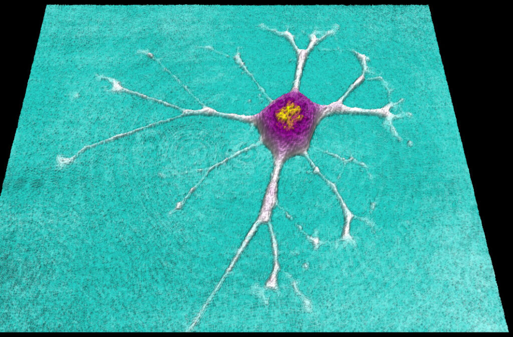
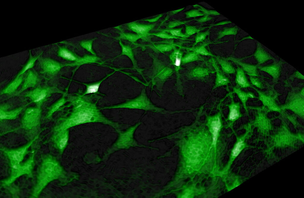
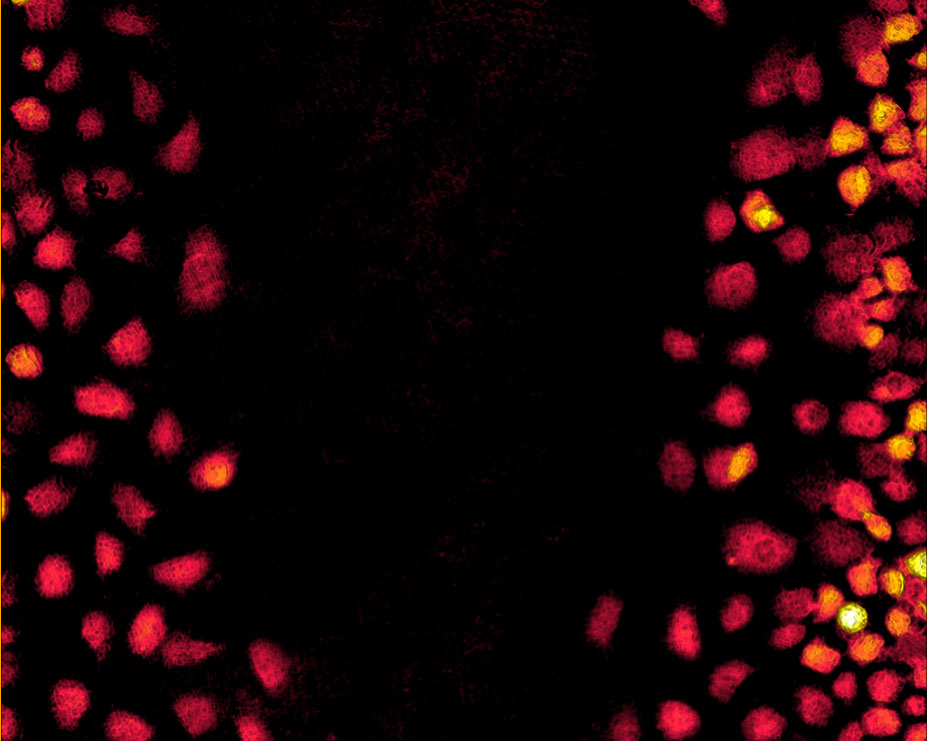
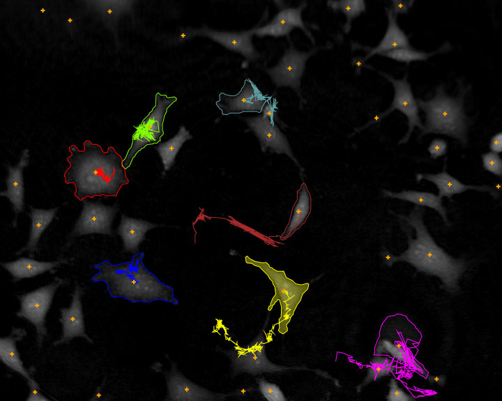
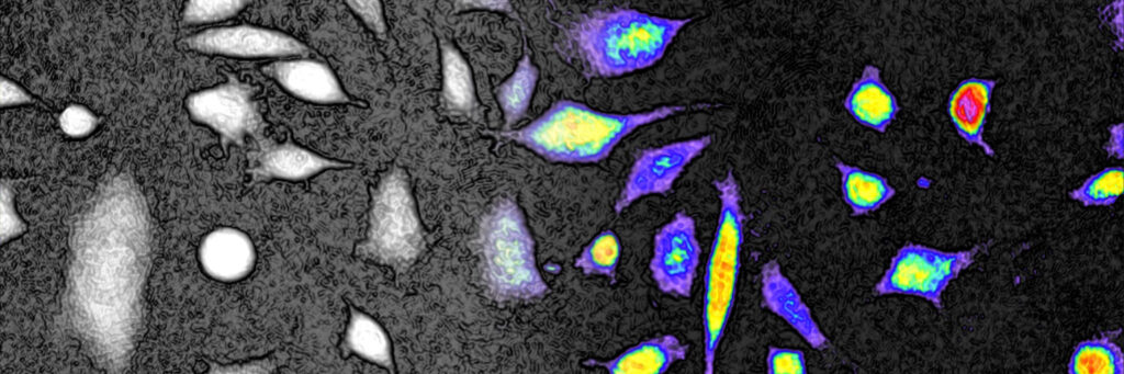
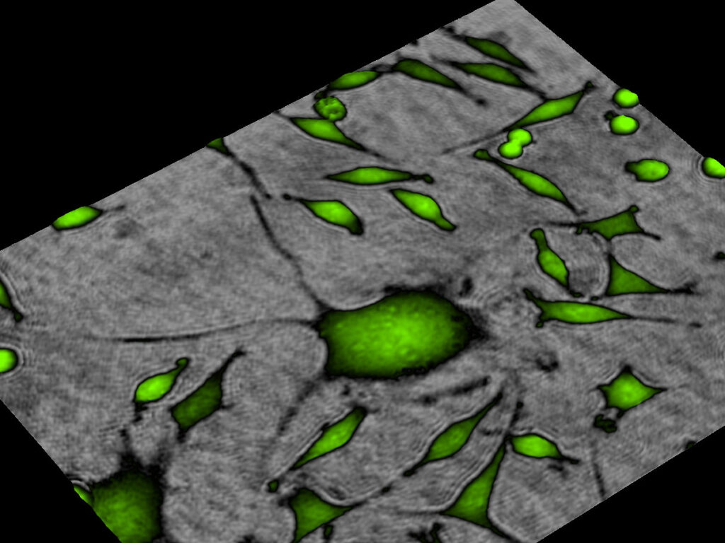
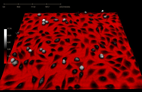
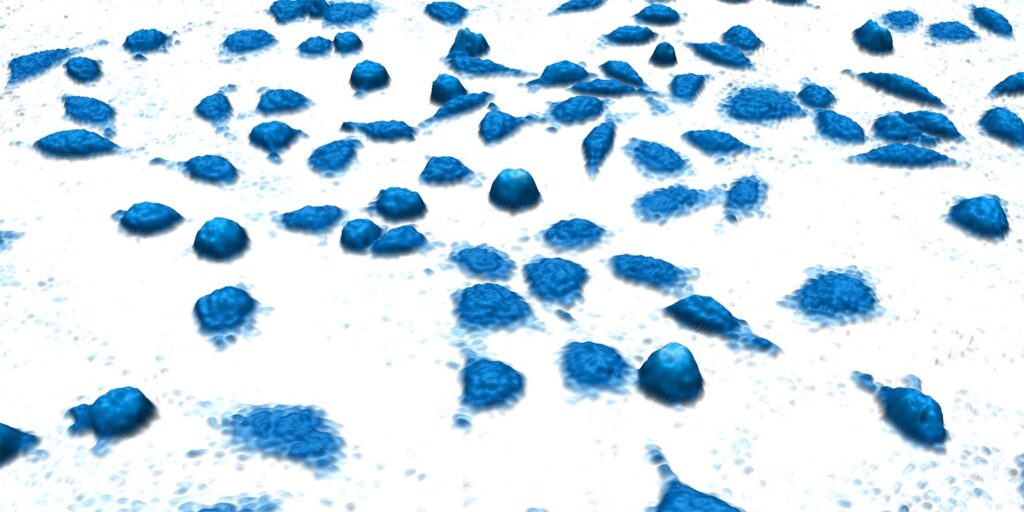
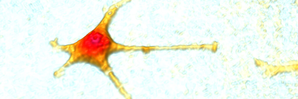
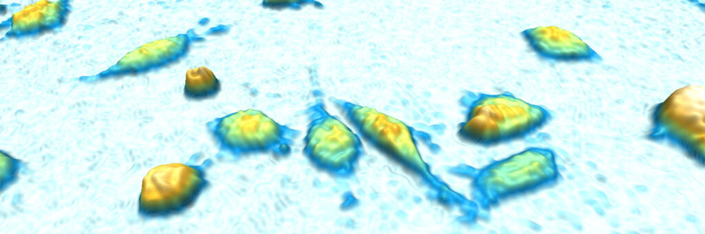
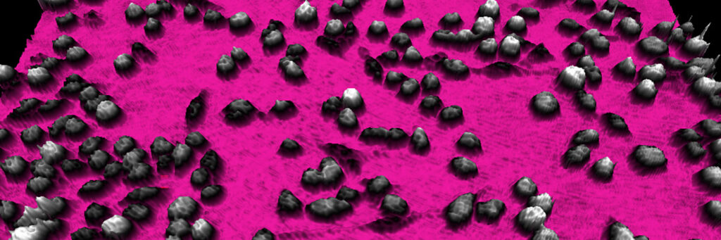
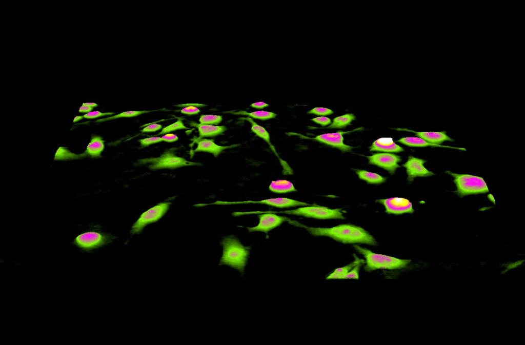
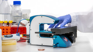
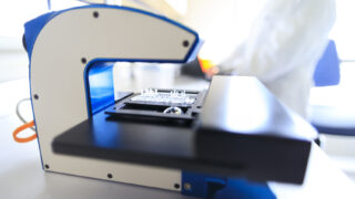
Researchers across the world employ HoloMonitor to automatically obtain live cell morphology, proliferation and migration data on a single-cell level, in real-time. HoloMonitor operates from within a standard cell incubator or hypoxia chamber to non-invasively provide cell-friendly cell analysis over extended periods.
Video Galleries

The PHI Story
Get to know PHI! PHI provides innovative tools that let you continuously image and monitor your cells directly in your incubator.
Learn more …
HoloMonitor How-to Tutorials
Learn how to handle the HoloMonitor Live Cell Imaging & Analysis System.
Learn more …
Introducing HoloMonitor M4
Get to know HoloMonitor M4 — an innovative, label-free live cell imaging and analysis system and its software App Suite!
Learn more …
Live Cell Imaging Videos
Live cell videos, imaged without labels, directly in an incubator by the HoloMonitor imaging system.
Learn more …
Never miss the latest news on label-free live cell imaging!
Follow us on social media!
Image Gallery
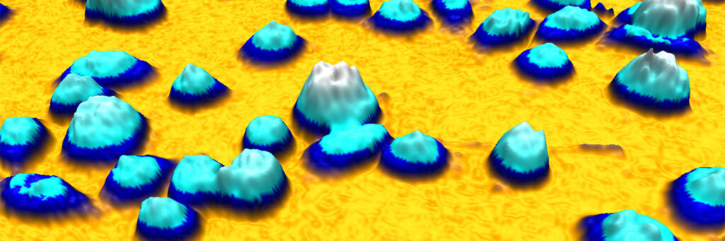
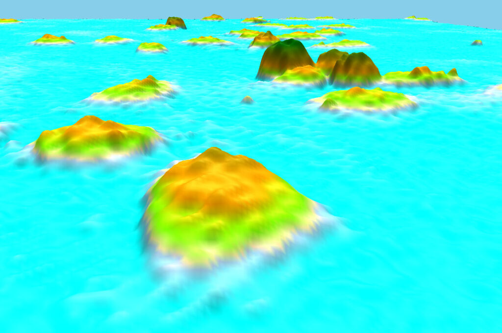
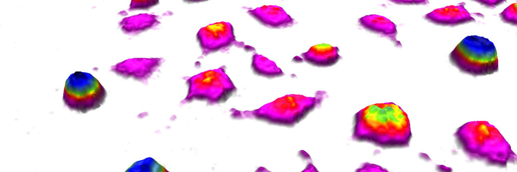
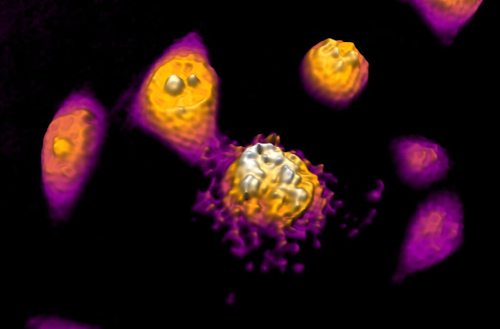
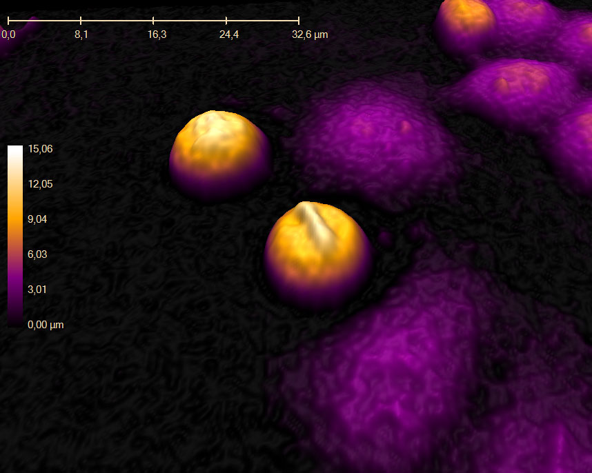
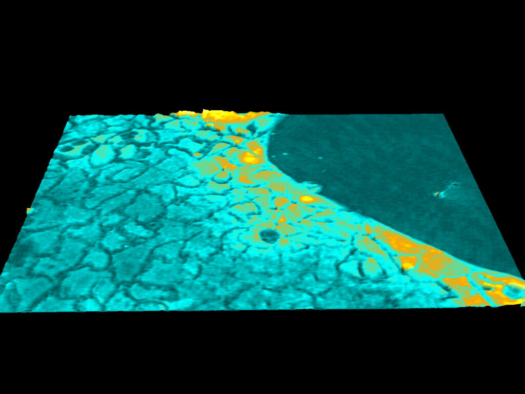
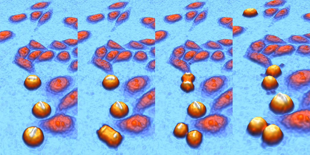
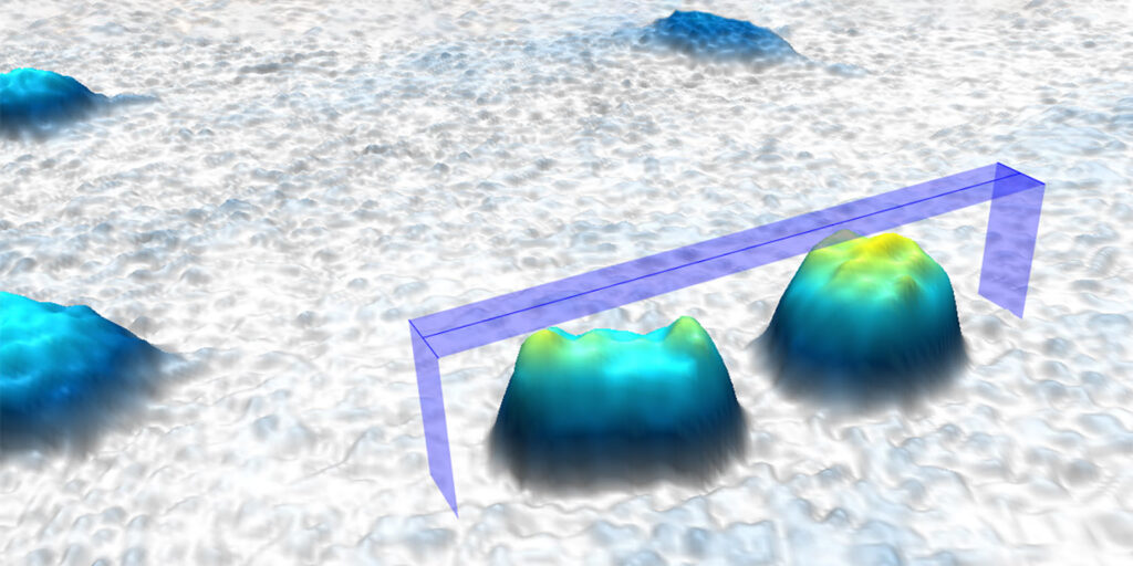
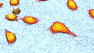
All HoloMonitor applications are label-free. The cell colors below are added by the HoloMonitor cell imaging software — App Suite.