2021 WSCS – A remarkable event

Over the last week, PHI had an excellent opportunity to attend the combined 2021 World Stem Cell Summit and Regenerative Medicine Essentials Course (WSCS). Our CSO, Kersti Alm, had an inspiring and educational talk about how HoloMonitor®, as an innovative technology, can facilitate stem cell-based regenerative medicine research.
The annual WSCS is the most inclusive and expansive conference in the stem cell and regenerative medicine fields. In addition, the summit provides a comprehensive look from the science behind leading discoveries to regulatory and manufacturing challenges within the field.
Indeed the regenerative medicine is an emerging field that involves repairing and regenerating body organs and tissues. Stem-cell-based cell therapy, in particular, is the rising star.
Generally, it involves introducing stem cell populations into patients to replace damaged tissues and cells. As a result, cell therapies may one day replace organ donation and eliminate the accompanying issues, such as rejection and tissue insufficiency.
In particular, researchers aim to cure common but severe diseases, such as diabetes, Parkinson’s, and several cancer forms.
Live Cell Imaging of Stem Cells
Label-free time-lapse video of differentiating human iPS cells.
Live cell imaging time-lapse of stem cells on microgrid arrays.
HoloMonitor at World Stem Cell Summit
In other words, cells are critical for regenerative medicine and stem cell research development. Thus, we have to study individual cells without reagents or other harmful effects in a controlled environment. In fact, HoloMonitor has such special capabilities and is well-suited for cell therapy methods in which patients are treated with laboratory-grown cells.
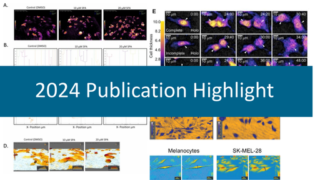
Our CSO, Kersti Alm, presented our cell-friendly live cell imaging microscope, HoloMonitor, and its application in stem cell research during the summit. The audience showed a great interest in HoloMonitor, especially in its label-free and imaging directly inside incubator features. For the past several years, scientists have markedly used HoloMonitor in different research areas. As a result of today, HoloMonitor is featured in more than 175 peer-reviewed scientific publications worldwide.
It was indeed an exciting summit to be at, where we had a fantastic opportunity to connect and share ideas with some of the most brilliant minds in regenerating medicine.
PHI CSO, kersti alm
What Did We Talked About?
In short, Kersti introduced the principle of digital holography imaging and why it is harmless to cells. Moreover, we discussed two application examples of HoloMonitor in stem cell research from our featured users.
Missed the talk? No worries, click below to bring you back to the summit.
To begin with, Patecchia and colleagues used HoloMonitor to investigate how late changes in the differentiation process affected their cells. In brief, they quantified skeletal stem cell trans-differentiation by measuring morphological parameters, particularly cell roughness and thickness. Therefore, it is possible to separate the undifferentiated stem cells from the fully differentiated adipocytes and osteoblasts.
On the other hand, H.E. Owston and colleagues investigated the “clinically accessible” samples of human periosteum, a highly vascular and multipotential stromal cell- (MSC-) rich tissue that is vital for fracture healing. In brief, this study aimed to compare human periosteum and donor-matched iliac crest bone marrow MSC content using HoloMonitor cell morphology and cell tracking assays. As a result, the unique morphological and migration characteristics of periosteal MSCs can be determined using the cell tracking assay, which is used to develop novel bone graft substitutes.
Wait, There Is More!
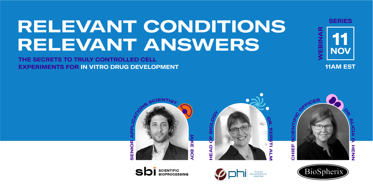
Our partners Scientific Bioprocessing (SBI) and BioSpherix, Ltd. have also participated in this fantastic summit. SBI develops real-time pH and dissolved oxygen sensors to monitor pericellular conditions. In addition, BioSpherix designs and builds cytocentric equipment for academic research, pharmaceutical and biotechnology laboratories around the world.
Remember that we held a joined three-part webinar series? Together with like-minded technology companies, we revealed our secrets to truly controlled cell experiments:
Series #3 – In vitro Drug Development
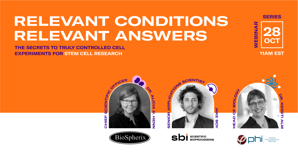
Series #2 – Stem Cell Research
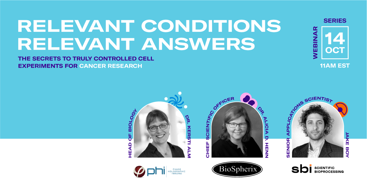
Series #1 – Cancer Research
References:
Petecchia et al. “A biophysical approach to quantify skeletal stem cells trans-differentiation as a model for the study of osteoporosis”. Biophysical Chemistry (2017).
Owston et al. “Colony Formation, Migratory, and Differentiation Characteristics of Multipotential Stromal Cells (MSCs) from “Clinically Accessible” Human Periosteum Compared to Donor-Matched Bone Marrow MSCs”. Stem Cells International (2019).