Picture-Perfect Science! HoloMonitor® Images Featured on Cover of Advanced Biology
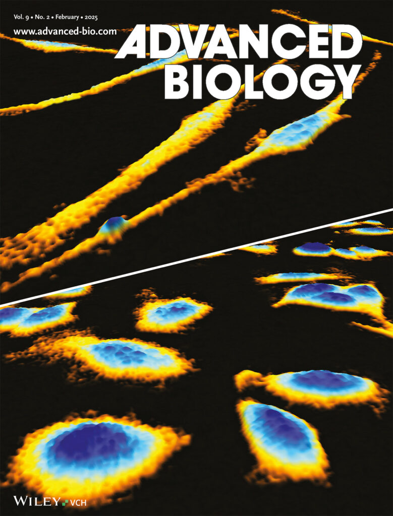
We are proud to announce that a recent study utilizing HoloMonitor® M4 has been featured on the cover of Advanced Biology, Volume 9, Issue 2 (February 2025). The study, led by Dr. Besa Xhabija and colleagues at the University of Michigan-Dearborn, USA, demonstrates the transformative potential of HoloMonitor, our non-invasive, real-time live cell imaging system, in advancing melanoma research.
The Study in Short
In this study, researchers used HoloMonitor to profile melanocytes and SK-MEL-28 melanoma cells with unprecedented precision, without the need for labels or any other damaging interventions.
Using advanced techniques, including Principal Component Analysis (PCA) and t-Distributed Stochastic Neighbor Emulation (t-SNE), the researchers discovered significant differences in cell size, shape, optical properties, and migration patterns between healthy melanocytes and malignant SK-MEL-28 melanoma cells.
The authors highlight these parameters as potential, non-invasive biomarkers for melanoma diagnostics and treatment.
Key Highlights of the Study
- Quantitative morphological differences: The study demonstrates distinct differences in cell area, volume, and diameter between normal melanocytes and SK-MEL-28 melanoma cells.
- Directional movement as a distinguishing parameter: Using the HoloMonitor single-cell tracking assay, the authors found that directional cell movement could serve as a distinguishing factor between melanocytes and SK-MEL-28 melanoma cells.
- Biomarker identification for diagnosis and therapy: The study emphasizes the potential of the identified parameters as biomarkers for cell characterization and differentiation, paving the way for more effective diagnostic and therapeutic strategies.
Featured on the Cover
HoloMonitor played a central role in advancing our melanoma research and contributed directly to our work being featured on the cover of Advanced Biology. I believe any biomedical research lab—especially those aiming to accelerate discovery efficiently—would benefit from using HoloMonitor as a powerful, non-invasive screening platform.”
Dr. Besa Xhabija
Assistant Professor, University of Michigan-Dearborn, USA
PHI congratulates Dr. Besa Xhabija and her research team on their groundbreaking work and for shining a spotlight on HoloMonitor and its role in melanoma research. Stay tuned for more updates as we continue to drive innovation in cellular imaging and cancer research!


