Cell Morphology Assay
Your non-invasive and high-content live cell morphology assay
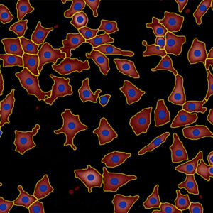
The label-free HoloMonitor® Cell Morphology Assay enables non-invasive live cell imaging and kinetic analysis of a wide range of morphological properties, including individual cell volume, area, and thickness. With the add-on fluorescence unit, you can add a green fluorescence dimension to the holography data, providing enhanced insights into your cell cultures.
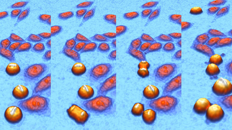
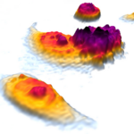
Time-lapse video showing the dynamic of living cells, captured with HoloMonitor.
Benefits
Why choose the HoloMonitor Cell Morphology Assay?
High-content
Study 30+ cell morphology features for every single cell in your population, plus extract proliferation & motility data.
Accurate
Get exact cell segmentation with digital holography to measure what you actually want to study.
Versatile
Detect early drug responses from cell morphology changes, study the cell cycle, analyze cell death kinetics or classify sub-populations. Applications are endless!
Continuous
Monitor cellular processes, cell behavior, and characteristics in real-time with a high temporal resolution. And as a result, you won’t miss any critical cellular events.
Assay Output
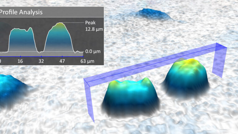
The HoloMonitor Cell Morphology Assay measures a wide range of morphological properties of individual cells, including cell volume, area, and thickness. The optional fluorescence dimension enables the integration of green fluorescence data into the label-free holography results.
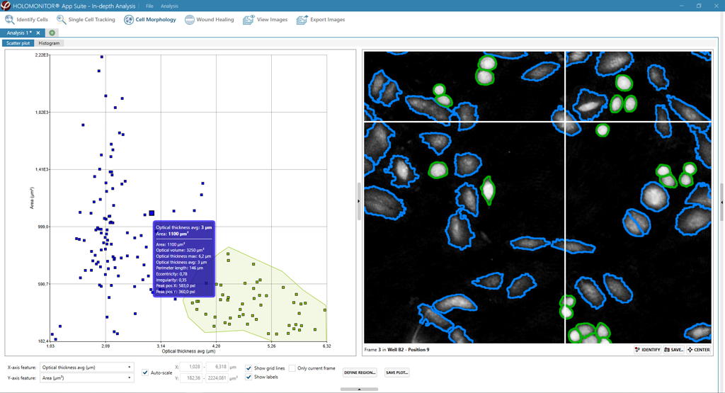
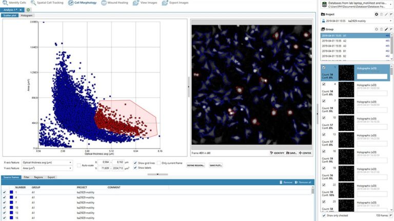
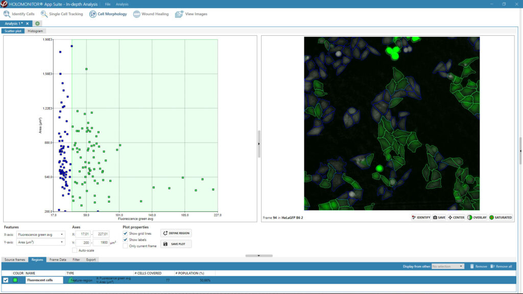
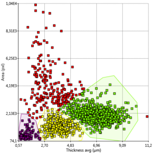
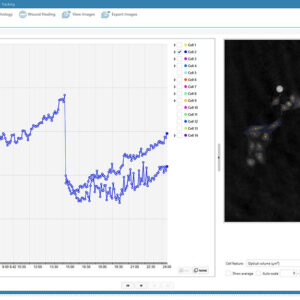
Scatter plots help visualize differences between control and treated cells. And they assist you in comparing cellular characteristics at different time points of the experiment.
Gating regions enable you to classify sub-populations and identify (rare) cells of interest. In addition, you could add HoloMonitor Single Cell Tracking for further single-cell analysis.
Moreover, histograms show the cell population distribution of selected morphological parameters at selected time points.

What results do you get with the HoloMonitor Kinetic Cell Morphology Assay?
- 30+ cell morphology features with the possibility for fluorescence
- Calculation spreadsheets of all data, including population averages
- Images and time-lapse videos of kinetic morphology changes
High-content.
Numerous results from one sample on single-cell level
Usually, we think of cell morphology as cell area, cell volume, and maybe cell shape. However, using HoloMonitor, you can analyze more than 30 different cell morphology features over time. Above all, many of those parameters are unique for our non-invasive digital holography technology. Now, this provides you with in-depth data on every single cell in your population. Furthermore, you can gain further results from the same sample by combining this assay with other HoloMonitor live cell assays or the add-on fluorescence unit. For example, you can extract cell proliferation and cell motility data for a more detailed picture.
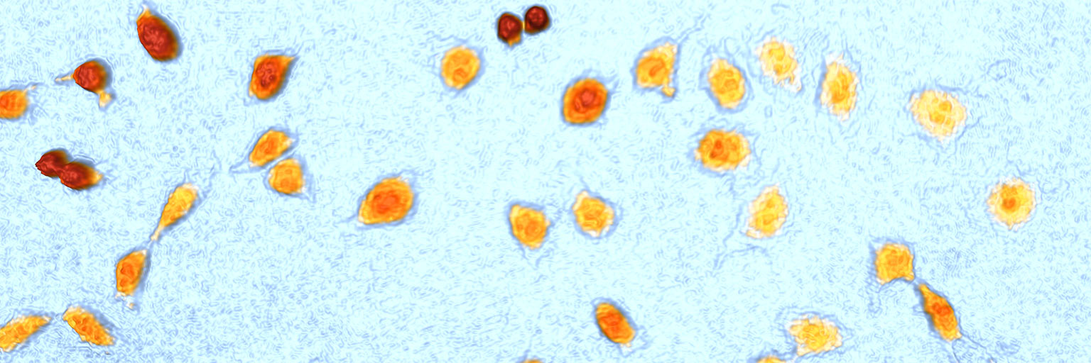
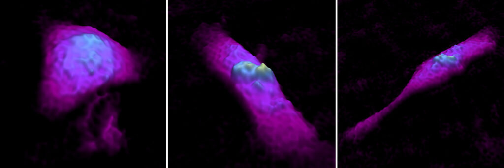
Same cell – many shapes!
Accurate.
Exact cell segmentation makes live cell identification simple
Firstly, individual living cells are much more easily singled out from the background with quantitative phase imaging in comparison to traditional methods, such as phase contrast. As a result, robust computer algorithms can automatically and precisely quantify cell morphology changes down to every single cell in your population. This way, you receive exact cell morphology assay results. And measure what you actually want to study.
On-demand Webinar
Watch our expert cell morphology webinar!
Join our PHI CSO, Kersti Alm, when she highlights the great potential of quantifying cell morphology, for both single-cell and cell populations.
Key Applications
What can you study with the HoloMonitor Cell Morphology Assay?
Cell life dynamics
Cell culture health & quality
Drug response & early drug effects
Cytotoxicity & cell death
Epithelial-mesenchymal transition (EMT)
Cell differentiation
Cell (state) classifications
Sub-population studies
Cell cycle studies
Cell division
Co-culture
Report gene expression
Uptake assay
Transfection efficiency
Versatile.
Kinetic cell morphology is in the focus of many applications
Many naturally occurring cell processes, such as cell division, cell density, and cell movement, affect cell morphology. Also, combining cell morphology assay results with cell proliferation and cell motility is a powerful tool. Not only, for example, for cytotoxicity studies or in vitro drug development.
Not only can you detect early drug responses from cell morphology changes. But also study the cell cycle, analyze cell death kinetics, or classify subpopulations.
Individual cell morphology insights are useful for:
- For evaluation of cell culture health and quality
- As early and sensitive markers to detect drug effects
- To provide information on drug mode of action
- For identification of dying cells
Cell Morphology Assay Protocol
How do you set up this in kinetic cell morphology assay?
1. Sample preparation
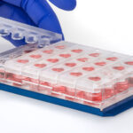
- Prepare your cell cultures in your standard vessel as you normally would.
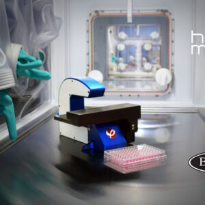
- Use HoloLids for optimal image quality and data acquisition.
- Click your cell culture vessel onto the HoloMonitor motorized stage and you are ready to go!
2. Assay setup
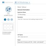
- Select the Cell Morphology Assay in the software and follow the guided workflow. Next, name your experiment and define treatments.
- Add the positions you want to image. Also, choose how often and for how long you want to image your cells.
- Start imaging and walk away!
3. Explore results
- Receive kinetic gap closure data and time-lapse videos in real-time. Analyze the results with other live cell assays for deeper insights into your cells.
- Export the raw data to Excel for further analysis and customized data presentation.
- Re-use the untouched cells for other experiments!
Continuous.
Never miss an important cellular event
HoloMonitor is designed to operate 24/7 directly inside your standard incubator. This makes it possible for you to continuously monitor and quantify your cell cultures in their undisturbed environment. The high temporal resolution of the system allows you to study in-depth cellular processes, cell behavior, and changing cell morphology characteristics in real-time without missing any important cellular events in your culture.
A panel of cellular parameters, including cell morphology, proliferation, and motility, were quantitatively measured and analyzed using [HoloMonitor]. This novel and quantitative approach provides the opportunity to continuously and non-invasively study relevant cellular processes, behaviors, and characteristics of cancer cells and other cell types in a simple and integrated manner.
Jing Huang et al.
A Time-lapse, Label-free, Quantitative Phase Imaging Study of Dormant and Active Human Cancer Cells, Journal of Visualized Experiments (2018)
Featured Publications
Browse our publication library for more references and see how our worldwide HoloMonitor users run their kinetic cell morphology analysis.
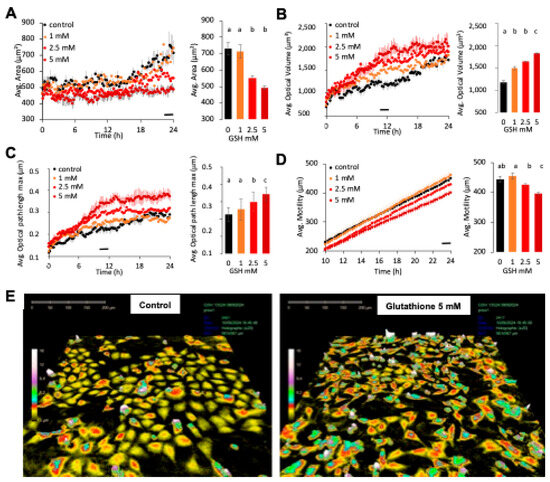
Comparison of Real-Time Methods Demonstrating the Effects of Reduced Glutathione on Olfactory Neuroblasts
Journal: Applied Sciences (2025)
Research Areas: Cell research
Cell Lines: 13s24
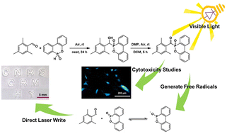
2,4,6-Trimethylbenzoyldiphenylphosphine oxide (TPO) analog: a non-cytotoxic type-I photoinitiator for free radical photopolymerization
Journal: Green Chemistry (2025)
Research Areas: Material science
Cell Lines: C3H10 T1/2
Cells Prioritize the Regulation of Cell Mass Density [not peer-reviewed]
Journal: bioRxiv (2024)
Research Areas: Cell Biology
Cell Lines: NIH3T3, HT1080, T47D, Hs578, RPE 2n, RPE 4n, MCF-10A, MDA-MB-231, and its single cell clones (SCCs) MDA-SCC-1006, MDA-SCC-1304, and MDA-SCC-1308

The Yin and Yang of hsa-miR-1244 expression levels during activation of the UPR control cell fate
Journal: Cell Communication and Signaling (2024)
Research Areas: Apoptosis Regulation
Cell Lines: HeLa S3, Calu3, 16HBE14o-