The PHI Blog
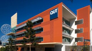
HoloMonitor down-under – Freya & Olaf advance QUT research
Freya and Olaf – two HoloMonitor systems – are expanding Queensland University of Technology’s Live Cell Imaging capabilities.
Learn more …
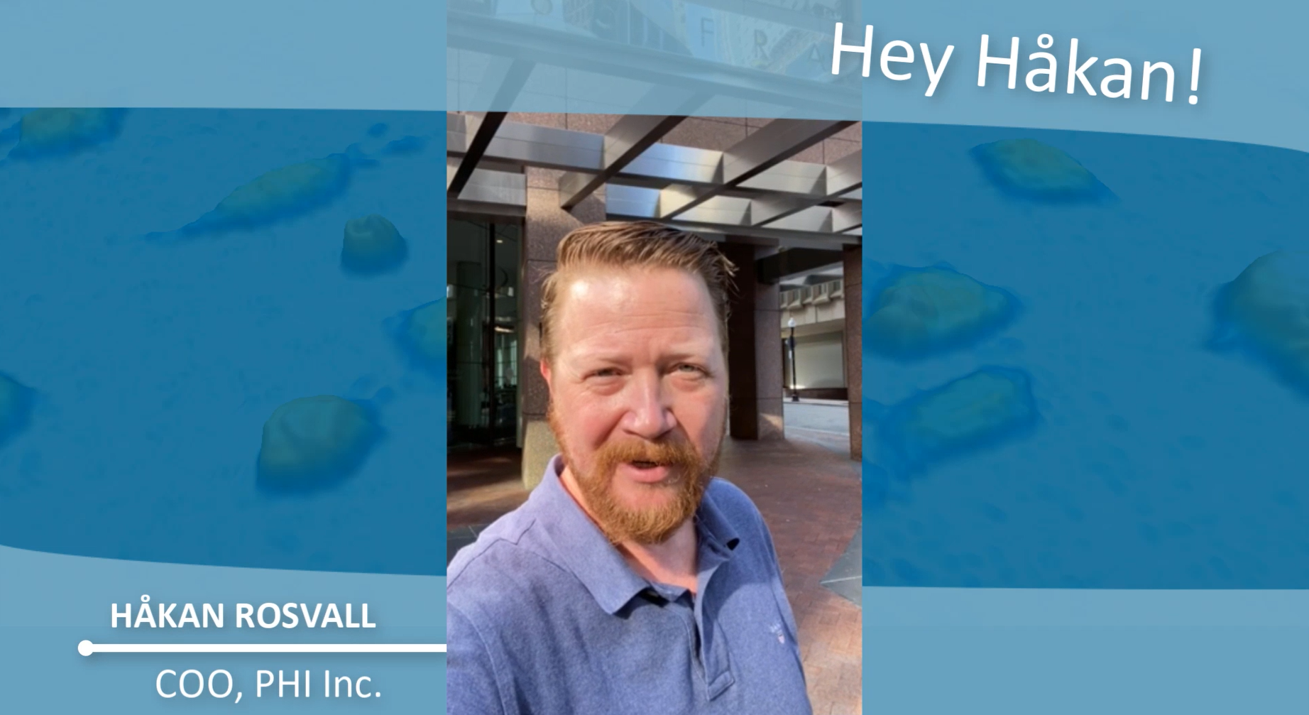
USA, Håkan is calling – pick up!
With the opening of the U.S. subsidiary we continue our global expansion. Greetings from Håkan Rosvall, Head of PHI Inc.!
Learn more …
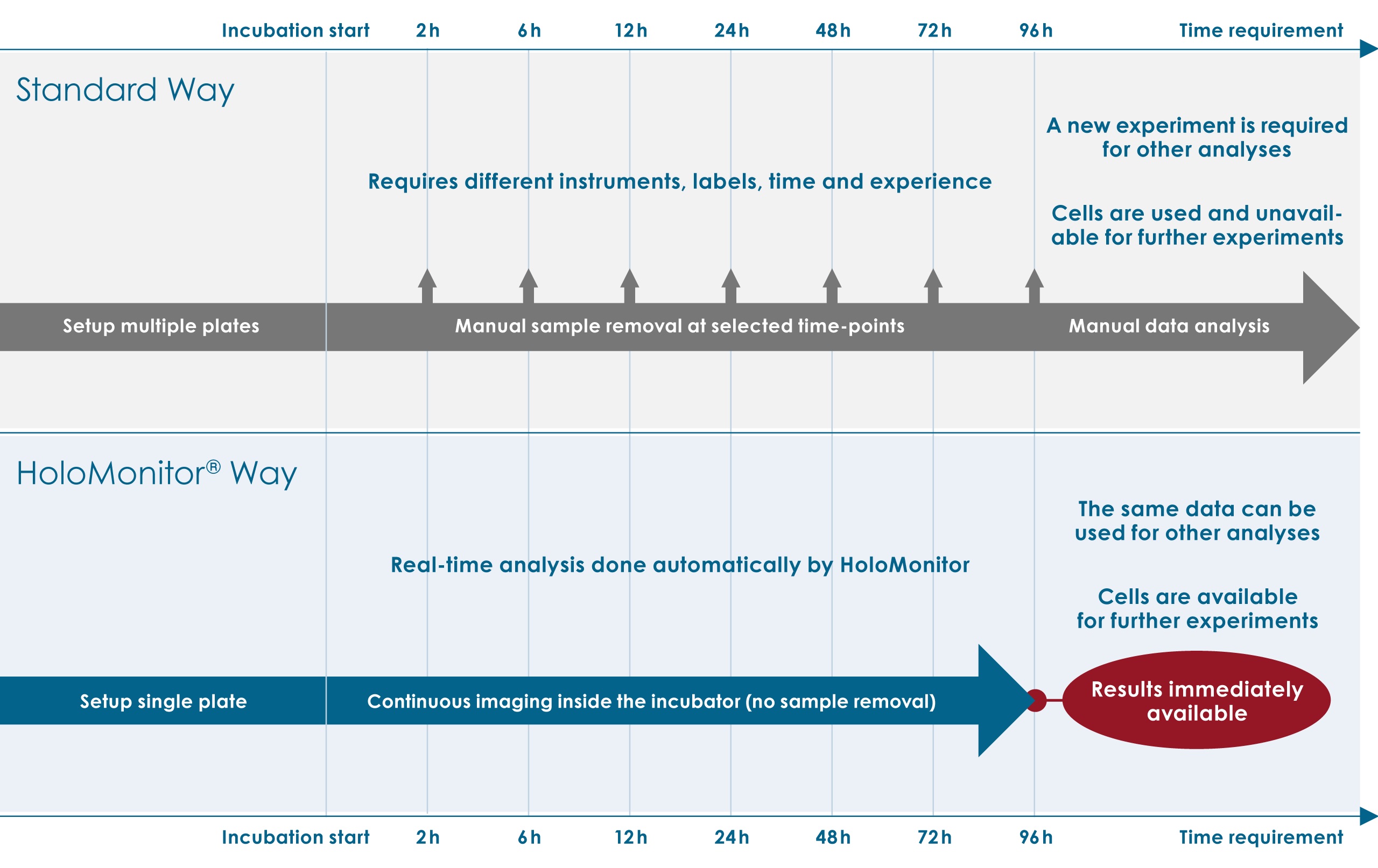
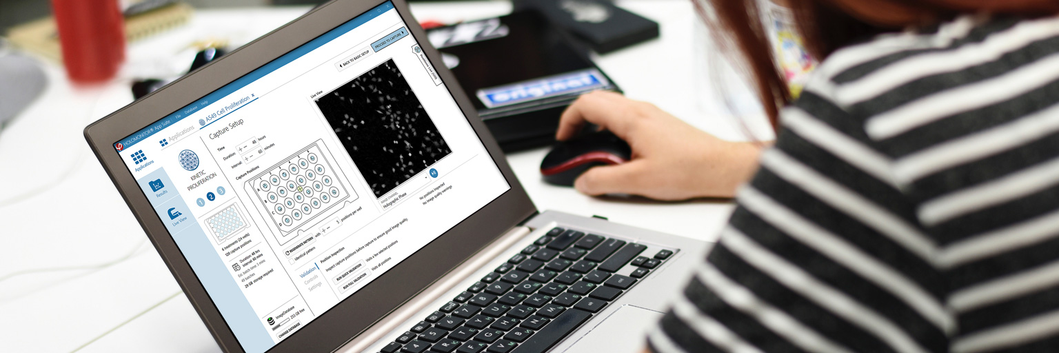
HoloMonitor saves your time, money – and cells
HoloMonitor® enables real-time label-free and quantitative studies of adherent cells directly in the cell incubator. Compared to standard methods the HoloMonitor approach saves both time, money and valuable cells for further experiments.
Learn more …
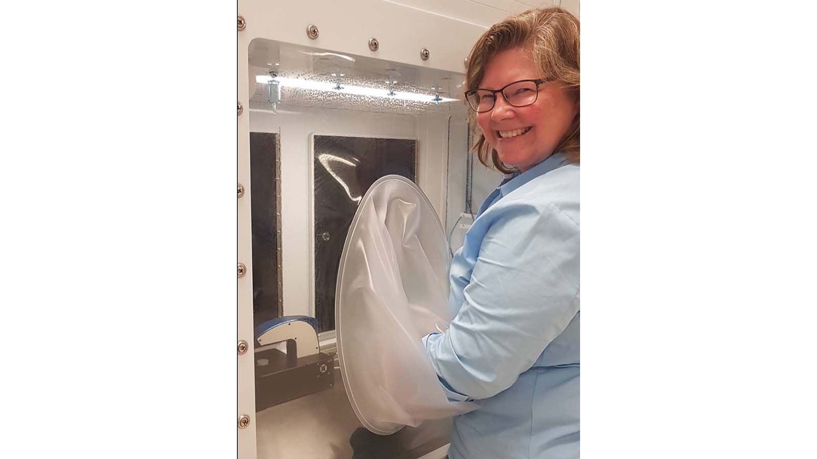
Growing & Analyzing Cells at their Best
BioSpherix and Phase Holographic Imaging (PHI) have teamed up to support researchers in their ambition to grow and analyze cells in a cell-friendly environment, without cell disturbing factors like labels, stains and temperature fluctuations.
Learn more …
Quantitative Live-Cell Imaging Analysis
Since 2014, HoloMonitor and quantitative live-cell imaging analysis play an important role in advancing the scientific interests of researchers at Northeastern University.
Learn more …
A study on Adipocyte Migration based on HoloMonitor single-cell level results
In yet another publication scientists report on how HoloMonitor was used to study cell motility and migration for deeper understanding of cell biological processes.
Learn more …