
Meet HoloMonitor®
A Cell-friendly Incubator Microscope
- Measure at once 30+ different cellular features from cell population down to single-cell level
- Quantitative cell imaging, directly in your incubator using standard cell culture vessels
- Easy and comprehensive presentation of real-time data and high-quality time-lapse videos
- Easy-to-use, guided software that lets you reanalyse your experiments for more results
A Proven Technology
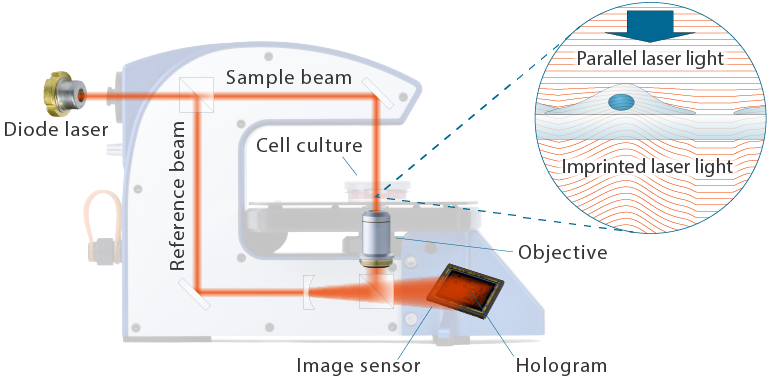
QPI imaging with digital holography
The label-free live cell imaging ability of HoloMonitor is based on the principle of quantitative phase imaging (QPI). Right inside your standard incubator, HoloMonitor enables intuitively non-invasive, real-time visualization and precise quantification of living cells without compromising cell integrity.
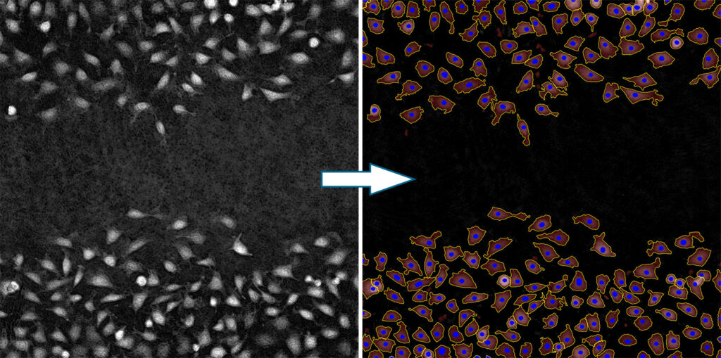
Simpler live cell identification
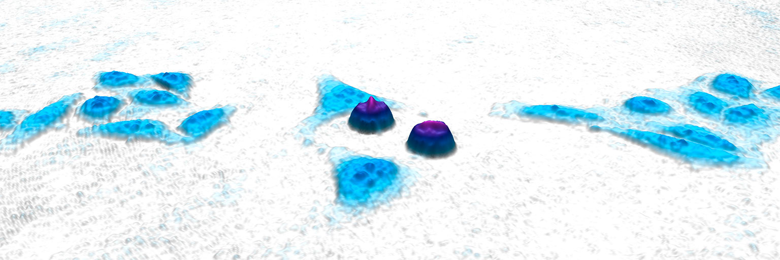
Your imaged cells appear as well-separated peaks from the background in a quantitative phase image. This way, HoloMonitor’s sophisticated software can automatically and robustly segment individual cells — of various confluencies, shapes and sizes.
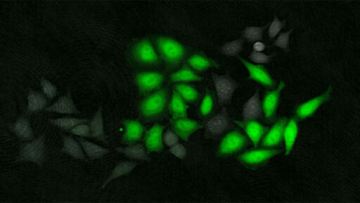
Additional insights with fluorescence
The latest HoloMonitor add-on fluorescence unit allows adding a green fluorescence dimension to the label-free holography data and studying your cells’ behavior in even more detail.
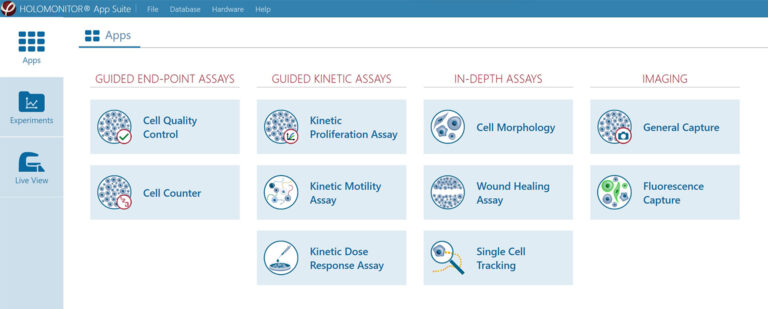
Applications
- Kinetic Cell Morphology
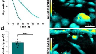
- Wound Healing (Scratch assay)
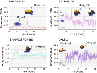
- Cell Division & Cell Cycle
- Cytotoxicity & Cell Death
- Reporter Gene Expression
- Transfection Efficiency
- Single Cell Tracking
- Cellular Differentiation
- Cell Proliferation
- Cell Counter
- Uptake Assay
- Live Cell Staining
- Chemotaxis
- Cell Motility & Migration
- Cell Growth
- Drug Dose-Response
- Cell Quality Control
- Co-Culture
Which application fits you most?
Find out how HoloMonitor can accelerate your research!
Multiple results from one experiment
Reanalyse your data with different HoloMonitor assays

The labs were still closed, and no additional experiments could be performed. Thus, the solutions had to come from our respective home offices. And here, the HoloMonitor’s built-in software solution showed its strength with a variety of applications and gave me the opportunity to re-analyze previous image material from home. New discoveries were made that strengthened our previous results.
Monica Hellesvik, Arnesen Lab, University of Bergen
Research from home blog post
New Application Notes
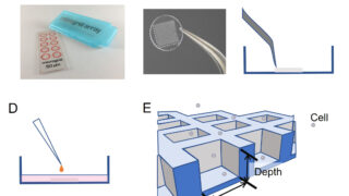
Using digital holography for imaging and analysis of cells in thin-layered 3D cultures
Quantitative long-term monitoring of non-adherent cells by digital holographic microscopy
Publications

KLF2+ stemness maintains human mesenchymal stem cells in bone regeneration
Journal: Stem Cells (2019)
Research Areas: Stem cell research
Cell Lines: hMSC, HUVEC
Digital Holographic Imaging as a Method for Quantitative, Live Cell Imaging of Drug Response to Novel Targeted Cancer Therapies
Journal: Theranostics: Methods and Protocols (2019)
Research Areas: Cancer research
Cell Lines: HeLa
Find out how other researchers use HoloMonitor:

Digital holographic microscopy: a noninvasive method to analyze the formation of spheroids
Journal: BioTechniques (2021)
Research Areas: Cancer research
Cell Lines: HT29
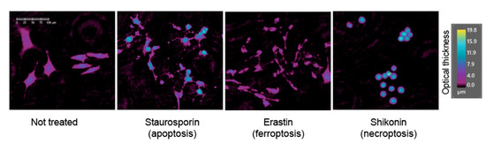
Label-Free Classification of Apoptosis, Ferroptosis and Necroptosis Using Digital Holographic Cytometry
Journal: Applied Sciences (2020)
Research Areas: Cell research
Cell Lines: 501mel
The HoloMonitor M4 is a beneficial device for live-cell imaging, providing a lot of data. The good thing is that you can analyze your experiment using one software assay, and later, you can rerun the recorded data in another assay, which allows you to get the maximum from your experiment. I like it and would recommend it to all people doing a cytotoxicity assessment based on cell proliferation and optical thickness of the adherent unstained cells.
Sasa Vasilijic, PhD
Stanford University